Next-Generation Sensors with Three-Dimensional Micro-/Nano-Structures
This is an Open Access article distributed under the terms of the Creative Commons Attribution Non-Commercial License(https://creativecommons.org/licenses/by-nc/3.0/) which permits unrestricted non-commercial use, distribution, and reproduction in any medium, provided the original work is properly cited.
Abstract
Innovative sensor technologies are crucial in various fields, including environmental monitoring, healthcare, and industrial applications. Sensor fabrication at the nano/microscale and the introduction of 3D structures can improve the sensitivity, response time, and overall performance of the sensor. Specifically, 3D architectures allow improved surface areas and interactions with target substances, whereas miniaturization facilitates improved sensitivity and integration into compact devices. This review discusses the recent advancements in five types of sensors, namely, gas sensors, strain sensors, environmental sensors, photodetectors, and biosensors, emphasizing the importance of structural innovations and size reduction in enhancing sensing capabilities. The essential design, fabrication methods, and potential applications are explored, highlighting the transformative impact of these technologies on future sensor development.
Keywords:
Micro/nano sensor, Three-dimensional (3D) sensor, Flexible sensor, Stretchable sensor, Soft electronic sensor1. INTRODUCTION
Sensor technology is essential for technological advancements in next-generation industries, such as environmental monitoring, healthcare, and smart cities. Sensors form the backbone of data collection and analysis, enabling real-time insights that enhance operational efficiency, safety, and sustainability. In addition, as industries increasingly seek solutions to complex challenges, the demand for more sensitive, accurate, and versatile sensors has intensified. This demand has prompted extensive studies on multidimensional structural design and miniaturization that can satisfy and exceed performance standards [1-5].
The introduction of three-dimensional (3D) micro- and nanoscale architectures into sensors offer unique advantages for the fabrication of next-generation high-performance sensors. 3D architectures can improve the sensor performance in various ways, including significantly increasing the surface area available for interaction with the specimen and enabling complex designs that facilitate better sensing mechanisms [6]. For example, an increased surface area can improve the sensitivity of gas sensors, as more active sites can interact with target molecules [7,8]. In addition, the unique physical and chemical properties of materials at the micro- and nanoscale, such as enhanced catalytic activity and tunable optical characteristics, are pivotal for the next generation of sensors [9,10]. Innovative designs that respond effectively to various stimuli can broaden the scope of sensor applications by manipulating their characteristics according to user requirements. Several advanced manufacturing techniques, such as simulation-based structural design and 3D printing, have been integrated with sensor fabrication technologies, enabling the fabrication of high-precision 3D micro- and nanostructures that were previously unattainable. Various manufacturing strategies can improve the manufacturability of complex architectures and create customized sensors that can adapt to specific environmental conditions or target analytes.
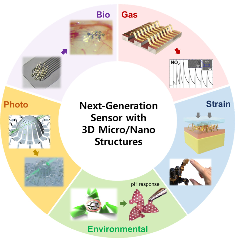
This review aims to provide a comprehensive understanding of the research that has introduced 3D micro- and nanoarchitectures into five key topics in the field of advanced sensors: 1) gas sensors [7], 2) strain sensors [8], 3) environmental sensors [9], 4) photosensors [10], and 5) biosensors [11], as shown in Fig. 1. Each section explores the recent advances, underlying principles, and potential applications based on various case studies, and explains how these technologies can be integrated into multiple fields, from industrial processes to personal health monitoring. For instance, gas sensors equipped with 3D nanostructures can detect minute amounts of analytes [7], whereas strain sensors can monitor body movements in real time [11]. Environmental sensors with multidimensional structures are vital for monitoring changes in ecosystems, and photosensors are crucial for tracking light paths in 3D spaces. In addition, miniaturized biosensors have the potential to revolutionize healthcare diagnostics by enabling the rapid and accurate detection of biomarkers [12,13]. Furthermore, through a comprehensive analysis of the current research and development, the innovative potential of 3D micro- and nanoscale architectures in the field of sensor technology are highlighted. A detailed review of these topics is expected to provide valuable insights into the ongoing evolution of sensor technology in terms of dimensions and scales as well as implications for future applications.
2. GAS SENSOR
In modern society, gas sensors play an essential role in ensuring the safety of various systems and the health of individuals. By enabling the early detection of harmful substances in the air, gas sensors contribute significantly to preventing accidents and protecting the environment, ultimately reducing loss of life and property damage. Additionally, they have been integrated into smart home systems and the Internet of Things, facilitating the effective monitoring and management of the surrounding environment. Users can monitor potential gas threats in real time through digital devices, such as smartphones, and receive immediate alerts in case of anomalies, thus further enhancing safety.
The structure and size of materials significantly influence the performance of gas sensors. Scaling down sensor materials can significantly increase the surface area and improve the sensitivity and response times. For example, the large surface area of nanoparticles increases the opportunities for contact with gas molecules, enabling faster and more accurate detection [14,15]. Furthermore, complex 3D structural designs can expand the contact area with gases and promote adsorption and reaction processes more effectively [16,17]. These innovations are crucial for maximizing the performance of gas sensors and establishing reliable safety systems for various environments. This chapter introduces various studies on improving the gas-sensor performance through miniaturization and structural design (Fig. 2).
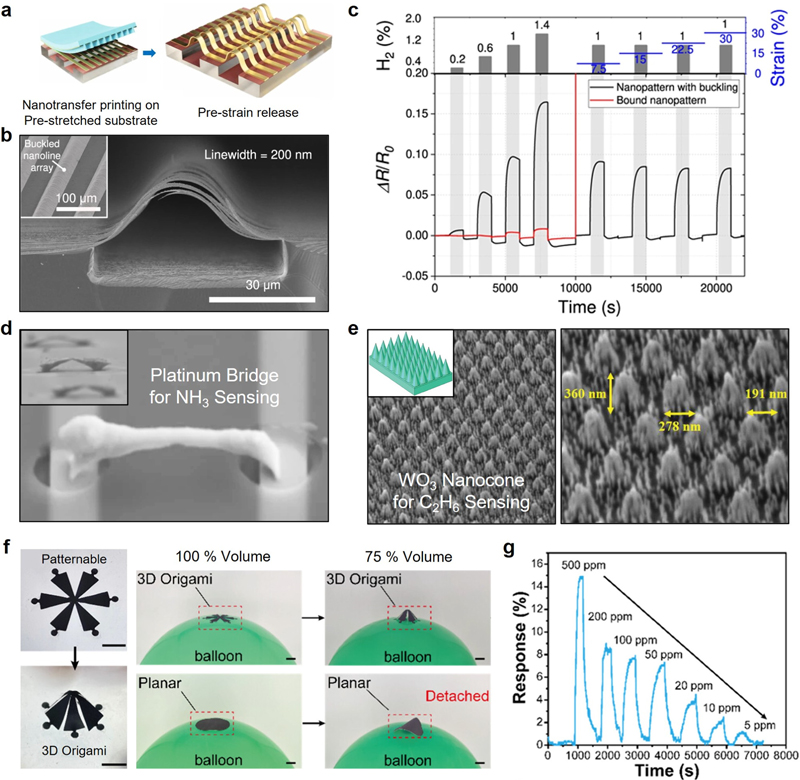
Highly sensitive gas sensors utilizing 3D micro/nanostructures to enhance the detection efficiency. (a) Fabrication process of 3D patterns using nanotransfer printing and buckling configuration. (b) Scanning electron microscope image of the buckled nanoline array. (c) Performance of flexible Pd-based hydrogen sensor under external strain. Ref. [7]; copyright [2023] Nature Publishing Group. (d) Array of self-aligning 3D nanobridges for ammonia gas sensing. Ref. [18]; copyright [2021] Nature Publishing Group. (e) 3D tungsten trioxide NC structure for ethane sensor. Ref. [8] Copyright [2020] John Wiley & Sons. (f) MXene/gelatin sensor electrodes with 3D origami structure. (g) Ammonia-sensing performance of 3D MXene origami sensor. Ref. [19] Copyright [2024] ACS Publications.
Ahn et al. demonstrated a method for fabricating nanoscale 3D structures based on a mechanical assembly, which allows the production of sensors that exhibit high sensitivity and maintain a stable performance under external strain [7]. In particular, two main aspects of 3D structure fabrication—design and manufacturing—have been the focus, enabling the creation of 3D nanostructures with diverse configurations and compositions. Using a covalent-bonding-assisted adhesive with a sub-10-nm thickness, two-dimensional (2D) nanotransfer printing on an elastomer substrate was achieved, resulting in 3D structures characterized by nanoscale-bound and suspended sites through a rational design and the prediction of the buckling configurations of the printed 2D patterns (Fig. 2 (a-b)). Furthermore, it has been validated that the stretchable Pd-based hydrogen sensor produced using this technology exhibits high sensitivity and retains a stable performance, even under external strain (Fig. 2 (c)). Isaac et al. reported a strategy for fabricating ammonia (NH3) gas sensors using a gas-phase electrodeposition method that can form an array of self-aligned 3D nanobridges (Fig. 2 (d)) [18]. Each 3D bridge structure provides a bias for gas sensing through parallel connections to an external circuit. Furthermore, a stable response of the NH3 gas sensor at various concentrations was achieved through an array structure containing 360 locally grown platinum bridges. Adib et al. proposed a highly sensitive ethane (C2H6) sensor based on a highly ordered 3D tungsten trioxide (WO3) nanocone (NC) structure (Fig. 2 (e)) [8]. The surface-to-volume ratio was enhanced by adopting the NC structure, allowing the metal oxide to interact with more reactants, thereby improving the sensitivity and performance of the gas sensor. In addition, simulation studies were conducted to determine the optimal design, and the crystallinity of WO3 and the NC structure were analyzed to maximize the sensor performance. Consequently, a superior sensing capability for C2H6 was demonstrated. Wang et al. reported a methodology for fabricating patterned electrodes using MXene/gelatin ink obtained through simple agitation and for fabricating 3D origami structures with the assistance of preprogrammed paper cutting and mechanically guided compressive buckling (Fig. 2 (f)) [19]. The sensors based on the 3D MXene origami structure stably adhered to the 3D curved surfaces, demonstrating a superior performance compared with their planar counterparts even under spatial changes. These 3D MXene origami sensors exhibited reactivity even to NH3 gas at 50 ppm, owing to the presence of rich surface functional groups (Fig. 2 (g)). Furthermore, the 3D structure successfully recognized the direction and height distribution of NH3 gas, indicating its potential to resolve the spatial distribution of toxic gases.
3. STRAIN SENSOR
Strain sensors measure the deformation or applied pressure of an object and are widely used in wearable devices and medical fields that require the monitoring of body movement and organ activity. In these applications, strain sensors enable the real-time detection of personal health status, facilitate disease prevention and early diagnosis, and prevent injuries by detecting the changes in strain during activities. In this respect, strain sensors have become essential technologies in various fields, such as healthcare, sports, robotics, and industrial monitoring, making significant contributions to enhancing individual safety and efficiency [20,21].
The performance of strain sensors is significantly affected by their structure and size. A 3D structure increases the surface area of the strain sensor, enhances the sensing sensitivity, and provides the flexibility required to adapt to various shapes and curved surfaces [22,23]. Accordingly, in fabricating strain sensors, the precise design of multidimensional structures is essential for improving performance and implementing functionality [24-26]. In this section, we introduce research on the introduction of 3D structures into strain sensors to maximize their performance and functionality and expand their applications (Fig. 3).
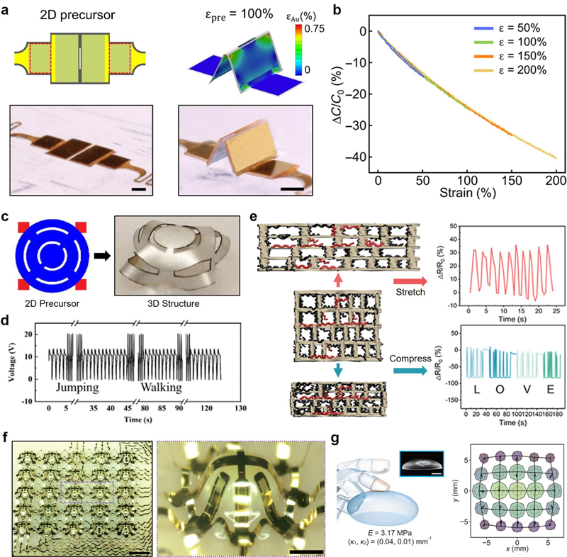
Strain sensors engineered with 3D micro/nanostructures for high sensitivity and flexibility. (a) Geometric transformation of the 2D precursors into origami-inspired 3D folded plates. (b) Sensing performance of the strain sensor under high strain rates. Ref. [27] Copyright [2023]. (c) PVDF piezoelectric thin-film pressure sensor with 3D Kirigami structures. (d) Plantar pressure sensing of the PVDF sensor with a 3D morphology. Ref. [28] Copyright [2024] Nature Publishing Group. (e) Foam strain sensor with stretchability and compressibility composed of styrene-ethylene-butylene-styrene and conductive CNTs. Ref. [29] Copyright [2022] ACS Publications. (f) Strain-sensing components arranged in 3D layout. (g) Tactile sensor system for elastic modulus and curvature sensing. Ref. [11] Copyright [2024].
Huang et al. reported a strain sensor with a 3D structure designed using morphologically deformable nonparallel plates [27]. The 3D strain sensor was implemented through the geometric transformation of a 2D precursor into an origamiinspired 3D folded plate (Fig. 3 (a)). The resulting strain sensor enabled large-angle deformations, achieving normal operation even at a high strain rate of 200% (Fig. 3 (b)) while maintaining high repeatability and stability for more than 700 cycles at 100% strain. Furthermore, the introduction of a unique 3D electrode design facilitated compressive strain sensing and directional strain responses, distinguishing it from existing strain sensors. The origami-inspired 3D stretchable strain sensor is expected to be a valuable strategy for applications in soft robotics and human–machine interfaces involving high strain rates. Zhang et al. developed a polyvinylidene fluoride (PVDF) piezoelectric thin-film pressure sensor featuring 3D Kirigami structures (Fig. 3 (c)) [28]. Based on experimental tests and finite element analysis, a fabrication strategy that allows the flexible tuning of the sensitivity and output voltage of the sensor by modifying the design of the 2D precursor and 3D architecture was proposed. The PVDF sensor with a 3D morphology successfully measured human pulse signals and plantar pressure and demonstrated exceptional operational stability under prolonged pressure conditions (Fig. 3 (d)). A strain sensor with a Kirigami-inspired 3D structure is expected to serve as a strategy to address the challenges associated with flexible and wearable pressure sensors. Guo et al. introduced a foam strain sensor that simultaneously exhibited stretchability and compressibility through the directional freeze drying of a composite material composed of conductive styrene-ethylene-butylene-styrene and conductive carbon nanotubes (CNTs), followed by surface coating with CNTs (Fig. 3 (e)) [29]. The foam strain sensor achieved a high stretchability of 250% and a compressibility of −50% while also attaining excellent elasticity and high orientation owing to its porous structure. The porous foam sensor demonstrated a stable signal output even under an ultrahigh strain of 800% and was capable of converting human gesture signals into Morse code or binary language. Owing to its excellent flexibility and stable electrical-mechanical properties, the foam-based strain sensor shows considerable potential as a wearable device for monitoring real-time human motions. Liu et al. reported bioinspired 3D architected electronic skin (3DAE-Skin), which mimics the spatial arrangement of Merkel cells and Ruffini endings in human skin [11]. The skin-like multilayer structure features force- and strain-sensing components arranged in a 3D layout, realized using lithographic techniques and precision mechanical assembly (Fig. 3 (f)). The 3DAE-Skin sensor exhibits excellent decoupled sensing performance for the normal force, shear force, and strain, enabling simultaneous measurements. Additionally, 3DAE-Skin integrates the data acquisition and processing modules, enabling tactile systems to measure the elastic modulus and curvature through simple touch (Fig. 3 (g)). This sensor shows potential for various bio-applications, such as food strength assessment and human–machine interaction.
4. ENVIRONMENTAL SENSOR
Modern societies face various environmental issues. Air pollution, water contamination, climate change, and biodiversity loss are serious threats to human health and ecosystems. Environmental sensors have become essential tools to address these problems. They detect and monitor pollutants in real time, contributing to policy decisions and public safety, while also helping establish early warning systems to minimize damage.
In this context, the miniaturization of environmental sensors facilitates easier installation and maintenance, allowing them to be deployed in diverse locations for improved data collection [30-32]. Additionally, 3D-structured environmental sensors can enhance sensitivity and enable precise measurements by increasing the surface area of the sensor [33,34]. Consequently, environmental sensors composed of miniaturized and 3D structured components are expected to be powerful tools for solving various environmental problems in modern society. This chapter introduces the research progress in these sensors (Fig. 4).
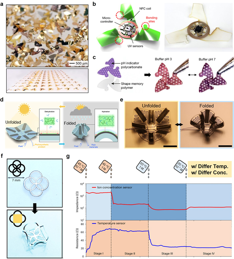
3D environmental sensors integrated with micro/nanostructures for multiple sensing capabilities. (a) Mesoscale 3D fliers inspired by wind-dispersed seeds. (b) Mechanically guided assembly of 3D mesostructures for wireless electronic devices. (c) Wireless pH and colorimetric sensor devices Ref. [35] Copyright [2021] Nature Publishing Group. (d) Illustration of environmentally responsive microfluidic devices inspired by plants. (e) Transformation of stimuli-responsive origami microfluidic device. Ref. [10] Copyright [2022]. (f) 3D structure formation owing to outward bending deformation. (g) Environmental sensors for changes in temperature and ion concentration. Ref. [36] Copyright [2024] Nature Publishing Group.
Kim et al. reported the fabrication of mesoscale 3D fliers inspired by wind-dispersed seeds, which provide unique capabilities for the aerial dispersal of advanced electronic devices (Fig. 4 (a)) [35]. A design that generates large momentum deficits in wakes induced by free fall promotes high drag forces and low terminal velocities, effectively eliminating instabilities in falling behavior and can be applied to millimeter-sized 3D structures. This technology has enabled the fabrication of miniature wireless electronic devices using a mechanically guided assembly of 3D mesostructures (Fig. 4 (b)). The resulting wireless devices operate without batteries and have potential applications in environmental monitoring, such as pH sensing and colorimetric sensors (Fig. 4 (c)). These biomimetic approaches are expected to provide important directions for the future advancement of microfliers and sensor technology. Pan et al. proposed an environmentally responsive microfluidic device inspired by plants with nastic movements in response to stimuli [10]. A transformable microfluidic chip was developed by integrating stimuli-responsive materials into a thin, foldable microfluidic chip. The entire device can be transformed according to temperature, humidity, and light irradiance (Fig. 4 (d-e)). This transformable origami microfluidic approach is named TransfOrigami microfluidics (TOM). In addition, TOM can be applied as an environmentally adaptive photomicroreactor, with transformations that reconstruct reaction channels and regulate photosynthetic conversion. Positive feedback control is embedded in the interactions between TOM and the environment, enhancing photosynthetic conversion when external conditions favor photoreactions. TOM will inspire energy, robotics, and biomedical applications that require environmental adaptation. Shin et al. developed a damage-free dry-transfer printing process for high-quality thin films using stress engineering, which enabled the fabrication of high-performance soft electronic devices [36]. By controlling the stress levels in the bilayer structure by adjusting the d.c. magnetron sputtering parameters and applying outward bending deformation to the mother substrate, the thin film was successfully peeled off from the substrate to create 3D structures (Fig. 4 (f)). Various patterns of 2D platinum films were transferred onto flexible substrates in either 2D or 3D forms using the proposed dry-transfer printing method, leading to the fabrication of an integrated sensor that combines ion concentration sensors and temperature sensors. The integrated sensor achieved successful environmental monitoring derived from structural changes owing to the temperature increases and impedance variations caused by differences in ion concentration (Fig. 4 (g)).
5. PHOTOSENSOR
Photosensors have attracted significant interest for monitoring in various devices owing to their advantages, such as high precision, reliability, fast response time, and noncontact operation. For example, they play a crucial role in environmental monitoring through real-time UV intensity and air pollution measurements and inpatient status monitoring via the noninvasive detection of biosignals. Photosensors ensure safety in autonomous vehicles by recognizing the driving environment. Thus, optical sensors serve as critical components in modern industries, such as environment, healthcare, and mobility.
When photosensors are fabricated at the nano- and microscales, they offer high sensitivity and fast response times. The small size increases the surface area of the sensor, making it more responsive to external light sources [37]. In addition, the introduction of a 3D structure enhances the light absorption efficiency and improves the sensor performance. The 3D structure also allows the utilization of various optical effects through complex shapes, contributing to the multifunctionality of the sensor [38,39]. Such modifications to the structure and size of photosensors expand their applicability across multiple industries and potentially drive further innovations. In this section, we discuss several research achievements that have increased the applicability of photosensors by introducing various sizes and structures (Fig. 5).
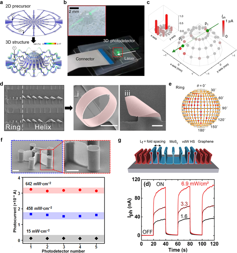
Photosensors designed with 3D micro/nanostructures to enhance the light detection performance. (a) Monolayer molybdenum disulfide and graphene-based origami-inspired 3D structures. (b) Transparent optical monitoring system with atomic-level thinness molybdenum disulfide. (c) Photosensor for light tracking in 3D space. Ref. [40] Copyright [2018] Nature Publishing Group. (d) 3D self-assembled structures of the Si/Cr rectangular nanomembranes. (e) 3D structured photodetector for detecting small incident light angles Ref. [9] Copyright [2024] Nature Publishing Group. (f) Template-assisted printed 3D star-shaped photodetector for light angle detection. Ref. [41] Copyright [2024] Springer. (g) Compressive-strain-induced 3D crumpled structure with high photoresponsivity. Ref. [42] Copyright [2020] Springer.
Lee et al. reported a 3D optical system for light detection and imaging by inducing the geometric transformation of 2D lightsensing materials incorporating monolayer molybdenum disulfide (MoS2) and graphene through compressive buckling [40]. A 3D array of MoS2/graphene photodetectors was fabricated by optimizing the assembly parameters for origami-inspired 3D shapes using finite element analysis. The set of assembly parameters enabled the implementation of the targeted 3D shapes from a 2D precursor through compressive buckling (Fig. 5 (a)). Consequently, atomic-level thin MoS2 served as the channel, whereas atomic-level thin graphene acted as the electrode supported on a polymer layer, forming a transparent optical monitoring system (Fig. 5 (b)). This system spontaneously forms a 3D configuration from a 2D planar geometry without the application of external forces, allowing the tracking of both the direction and intensity of the incident light in 3D space (Fig. 5 (c)). Zhang et al. introduced a quasi-static multilevel finite element method that emphasizes the geometry and boundary conditions of nanomembranes to fabricate 3D self-assembled structures [9]. The self-assembly mechanism was studied using rectangular Si/Cr nanomembranes, and the elastic energy changes were quantitatively simulated to successfully produce large-scale 3D structures of various shapes (Fig. 5 (d)). This manufacturing approach reproduced anisotropy across multiple material systems and pattern sizes, thereby demonstrating its excellent generalizability. Accordingly, various 3D structure photodetectors with an accuracy of 10° for incident light angle detection have been developed, showing potential for manufacturing electronic and photoelectronic devices (Fig. 5 (e)). Pan et al. fabricated a 3D star-like photodetector using a template-assisted printing strategy (Fig. 5 (f)) [41]. Template-assisted printing guides the polymer droplets to form a subwavelength structure, producing a star-like arrangement. The star-like 3D structure enabled light angle detection with resolutions of 10° in the vertical space and 36° in the horizontal space. In addition, 3D geometry offers enhanced spatial detection compared with conventional sensors. Furthermore, the optical resonance effect of the subwavelength façade and the shielding effect of the spatial arrangement facilitates the precise detection of light with weak intensity. This study is significant as it provides a novel prototype for 3D detection systems. Hossain et al. presented a methodology for fabricating optoelectronic devices based on crumpled 2D materials [42]. A van der Waals heterostructure composed of transition metal dichalcogenides and graphene was exploited, and this structure formed a 3D crumpled shape through compressive strain (Fig. 5 (g)). A 3D structure can accommodate significant stress through mechanical deformation, which makes it significantly more deformable than conventional thin films. Additionally, the optoelectronic properties of the crumpled heterostructure were not significantly affected by deformation, exhibiting photoresponsivity under varying light-intensity conditions. This fabrication method can be applied to various 2D heterostructured devices, creating possibilities for crumpled circuitry.
6. BIOSENSOR
Biosensors are essential for managing individual health, maintaining public health, and improving the quality of life. In particular, during the COVID-19 pandemic, biosensors have gained recognition for their contribution to public health protection through the early diagnosis and monitoring of infectious diseases. Furthermore, there is considerable anticipation for integrating biosensors with healthcare technologies as they can provide real-time health monitoring and personalized medical solutions [43-46]. Consequently, biosensors have become essential for addressing various healthcare challenges in modern society.
The introduction of nano- and micro-sized structures in biosensor fabrication enables high sensitivity and rapid response time [47]. Smaller dimensions increase the surface area of the sensor, making interactions with target molecules easier. Additionally, implementing a 3D structure expands the surface area of the sensor, thereby increasing its contact area with various biological samples [48]. This enabled the capture of more analytes, thereby enhancing the performance of the sensor. In addition, a 3D structure improves the detection capabilities in complex biological environments and provides the potential for integrating multiple functions. These structural enhancements significantly improved the accuracy and efficiency of the biosensor (Fig. 6).
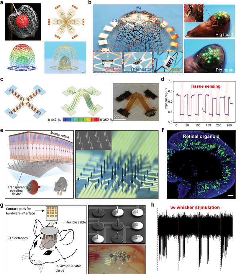
Biomedical sensors incorporating 3D micro/nanostructures for precise and efficient biosignal monitoring. (a) 3D microlattice structure formed using machine-learning-based shape programming. (b) Adaptive cardiac electronics and 3D electronic cell scaffolds with a 3D microlattice structure. Ref. [49] Copyright [2023]. (c) Flexible WBG electronics with complex 3D mesostructures. (d) Cell impedance change measurement of WBG electronics. Ref. [50] Copyright [2023] John Wiley & Sons. (e) 3D columnar flexible microelectrode with biocompatible liquid metal. (f) Electrophysiological monitoring of retinal organoids. Ref. [12] Copyright [2024] John Wiley & Sons. (g) High-aspect-ratio 3D MEA for monitoring 3D neuronal systems (h) Whisker stimulation with high signal-to-noise ratio. Ref. [13] Copyright [2024] John Wiley & Sons.
Cheng et al. introduced a microlattice design that transformed 2D films into programmable 3D curved mesosurfaces through a mechanically guided assembly. Analytical modeling and a machine-learning-based approach were employed for shape programming, leading to diverse 3D geometries (Fig. 6 (a)) [49]. Approximately 30 geometries were presented, including both regular and biological mesosurfaces. This microlattice design can be applied to a wide range of materials, such as silicon, metals, chitosan, and polyimide, providing higher feasibility than previous local stiffness control strategies. The 3D structures offer significant deformation capabilities and are more flexible than conventional thin films. Furthermore, these structures can be utilized in conformal cardiac electronic devices and 3D electronic cell scaffolds (Fig. 6 (b)). This microlattice structure has significant potential for enhancing the performance and applicability of biosensors. Truong et al. reported a novel engineering approach to create soft 3D mesostructured wide bandgap (WBG) materials without sacrificial layers [50]. This method contributes to the transformation of 2D precursors into complex 3D mesostructures through mechanical buckling and enables the fabrication of scalable and flexible WBG electronic devices on standard Si platforms without relying on traditional mechanical stamping processes (Fig. 6 (c)). 3D structures offer the advantages of stacking and patterning various materials based on their mechanical robustness and optical transparency. The diverse 2D and 3D architectures are equipped with mechanical, thermal, and impedance-sensing functionalities, providing promising opportunities for biosensor applications, including the impedance measurements of cells (Fig. 6 (d)). Lee et al. presented a strategy for fabricating soft microelectrodes with a 3D pillar shape that could record signals in retinal organoids using a biocompatible liquid metal (Fig. 6 (e)) [12]. The flexibility and delicate structure of 3D liquid-metal electrodes enable minimal invasion into the tissue, making them suitable for the long-term monitoring of neural activity. Liquid metal possesses a Young’s modulus similar to that of the tissue, which helps prevent chronic damage from rigid materials. In addition, microelectrodes can be positioned precisely on specific cell layers to collect electrophysiological data. The electrophysiological monitoring of retinal organoids using liquid-metal electrodes is feasible and contributes to the understanding of retinal functionality and synaptic connectivity (Fig. 6 (f)). This study demonstrates that 3D liquid-metal microelectrodes enable stable live-cell recordings, suggesting potential biosensor applications. Shihada et al. reported a simple and customizable 3D microelectrode array (MEA) fabrication process that enabled the manufacture of high-aspect-ratio 3D microelectrodes for investigating complex 3D neuronal systems (Fig. 6 (g)) [13]. A 3D lithography tool was used to print hollow pillars onto various planar MEA substrates, with these pillars serving as templates to guide the electrochemical deposition of conductive materials, allowing the growth of 2D electrodes into 3D. The 3D structure demonstrates flexibility and reproducibility during the fabrication process, enabling the production of 3D MEAs on both stiff and flexible MEA substrates. This versatility allows the electrochemical fabrication of 3D microelectrodes, which can be easily adapted to different electrode geometries when integrated with CMOS technology. 3D MEA technology records neural activity, such as whisker stimulation, with high signal-to-noise ratios and has potential applications across various model systems (Fig. 6 (h)).
7. CONCLUSIONS
This review highlighted the importance of 3D micro/nanostructures in advanced sensor technologies, focusing on gas sensors, strain sensors, environmental sensors, photosensors, and biosensors. We explored the fabrication strategies used to integrate 3D structures and micro/nanostructures into sensor technologies and discussed how these structures enhance sensor performance and sensitivity. We also discussed the recent innovations in sensor applications, including environmental change detection, biological process monitoring, and real-time data acquisition. The adoption of 3D micro-/nanostructures enhances the functional capabilities of these sensors and expands their potential for integration into complex systems. Developing efficient fabrication techniques for these complex structures is essential for maximizing their applicability in various fields. These advances demonstrate the innovative potential of 3D micro/nanostructured sensors in engineering and are expected to pave the way for novel solutions for practical applications in the near future.
Acknowledgments
This study was supported by the National Research Foundation of Korea (NRF) grant funded by the Korean Government (MSIT) (No. RS-2024-00347619).
REFERENCES
-
C. Dai, Z. Lin, K. Agarwal, C. Mikhael, A. Aich, K. Gupta, and J.-H. Cho, “Self-assembled 3D nanosplit rings for plasmon-enhanced optofluidic sensing”, Nano Lett., Vol. 20, No. 9, pp. 6697-6705, 2020.
[https://doi.org/10.1021/acs.nanolett.0c02575]

-
Q. Wang, Z. Yao, C. Zhang, H. Song, H. Ding, B. Li, S. Niu, X. Huang, C. Chen, and Z. Han, “A Selective-Response Hypersensitive Bio-Inspired Strain Sensor Enabled by Hysteresis Effect and Parallel Through-Slits Structures”, Nano Micro Lett., Vol. 16, No. 1, p. 26, 2024.
[https://doi.org/10.1007/s40820-023-01250-y]

-
Y. Gu, C. Wang, N. Kim, J. Zhang, T. M. Wang, J. Stowe, R. Nasiri, J. Li, D. Zhang, and A. Yang, “Three-dimensional transistor arrays for intra-and inter-cellular recording”, Nat. Nanotechnol., Vol. 17, pp. 292-300, 2022.
[https://doi.org/10.1038/s41565-021-01040-w]

-
J. Hu, Y. Qiu, X. Wang, L. Jiang, X. Lu, M. Li, Z. Wang, K. Pang, Y. Tian, and W. Zhang, “Flexible six-dimensional force sensor inspired by the tenon-and-mortise structure of ancient Chinese architecture for orthodontics”, Nano Energy, Vol. 96, p. 107073, 2022.
[https://doi.org/10.1016/j.nanoen.2022.107073]

-
B. W. Blankenship, Z. Jones, N. Zhao, H. Singh, A. Sarkar, R. Li, E. Suh, A. Chen, C. P. Grigoropoulos, and A. Ajoy, “Complex Three-Dimensional Microscale Structures for Quantum Sensing Applications”, Nano Lett., Vol. 23, No. 20, pp. 9272-9279, 2023.
[https://doi.org/10.1021/acs.nanolett.3c02251]

-
Q. Liu, W. Wang, M. F. Reynolds, M. C. Cao, M. Z. Miskin, T. A. Arias, D. A. Muller, P. L. McEuen, and I. Cohen, “Micrometer-sized electrically programmable shape-memory actuators for low-power microrobotics”, Sci. Rob., Vol. 6, No. 52, p. eabe6663, 2021.
[https://doi.org/10.1126/scirobotics.abe6663]

-
J. Ahn, J.-H. Ha, Y. Jeong, Y. Jung, J. Choi, J. Gu, S. H. Hwang, M. Kang, J. Ko, and S. Cho, “Nanoscale three-dimensional fabrication based on mechanically guided assembly”, Nat. Commun., Vol. 14, p. 833, 2023.
[https://doi.org/10.1038/s41467-023-36302-9]

-
M. R. Adib, V. V. Kondalkar, and K. Lee, “Development of highly sensitive ethane gas sensor based on 3D WO3 nanocone structure integrated with low-powered in-plane microheater and temperature sensor”, Adv. Mater. Technol., Vol. 5, No. 5, p. 2000009, 2020.
[https://doi.org/10.1002/admt.202000009]

-
Z. Zhang, B. Wu, Y. Wang, T. Cai, M. Ma, C. You, C. Liu, G. Jiang, Y. Hu, and X. Li, “Multilevel design and construction in nanomembrane rolling for three-dimensional angle-sensitive photodetection”, Nat. Commun., Vol. 15, p. 3066, 2024.
[https://doi.org/10.1038/s41467-024-47405-2]

-
Y. Pan, Z. Yang, C. Li, S. U. Hassan, and H. C. Shum, “Plant-inspired TransfOrigami microfluidics”, Sci. Adv., Vol. 8, No. 18, p. eabo1719, 2022.
[https://doi.org/10.1126/sciadv.abo1719]

-
Z. Liu, X. Hu, R. Bo, Y. Yang, X. Cheng, W. Pang, Q. Liu, Y. Wang, S. Wang, and S. Xu, “A three-dimensionally architected electronic skin mimicking human mechanosensation”, Science, Vol. 384, No. 6699, pp. 987-994, 2024.
[https://doi.org/10.1126/science.adk5556]

-
S. Lee, W. G. Chung, H. Jeong, G. Cui, E. Kim, J. A. Lim, H. Seo, Y. W. Kwon, S. H. Byeon, and J. Lee, “Electrophysiological Analysis of Retinal Organoid Development Using Three-Dimensional Microelectrodes of Liquid Metals”, Adv. Mater., Vol. 36, No. 35, p. 2404428, 2024.
[https://doi.org/10.1002/adma.202404428]

-
J. Abu Shihada, M. Jung, S. Decke, L. Koschinski, S. Musall, V. Rincón Montes, and A. Offenhäusser, “Highly Customizable 3D Microelectrode Arrays for In Vitro and In Vivo Neuronal Tissue Recordings”, Adv. Sci., Vol. 11, No. 13, p. 2305944, 2024.
[https://doi.org/10.1002/advs.202305944]

-
M. Guo, N. Luo, Y. Chen, Y. Fan, X. Wang, and J. Xu, “Fast-response MEMS xylene gas sensor based on CuO/WO3 hierarchical structure”, J. Hazard. Mater., Vol. 429, p. 127471, 2022.
[https://doi.org/10.1016/j.jhazmat.2021.127471]

-
B. Sun, F. Qin, L. Jiang, J. Gao, Z. Liu, J. Wang, Y. Zhang, J. Fan, K. Kan, and K. Shi, “Room-temperature gas sensors based on three-dimensional Co3O4/Al2O3@Ti3C2Tx MXene nanocomposite for highly sensitive NOx detection”, Sens. Actuator B Chem., Vol. 368, p. 132206, 2022.
[https://doi.org/10.1016/j.snb.2022.132206]

-
W. Tang, Z. Chen, Z. Song, C. Wang, Z. Wan, C. L. J. Chan, Z. Chen, W. Ye, and Z. Fan, “Microheater integrated nanotube array gas sensor for parts-per-trillion level gas detection and single sensor-based gas discrimination”, ACS Nano, Vol. 16, No. 7, pp. 10968-10978, 2022
[https://doi.org/10.1021/acsnano.2c03372]

-
Z. Yang, S. Lv, Y. Zhang, J. Wang, L. Jiang, X. Jia, C. Wang, X. Yan, P. Sun, and Y. Duan, “Self-assembly 3D porous crumpled MXene spheres as efficient gas and pressure sensing material for transient all-MXene sensors”, Nano Micro Lett., Vol. 14, p. 56, 2022.
[https://doi.org/10.1007/s40820-022-00796-7]

-
N. A. Isaac, J. Reiprich, L. Schlag, P. H. Moreira, M. Baloochi, V. A. Raheja, A.-L. Hess, L. F. Centeno, G. Ecke, and J. Pezoldt, “Three-dimensional platinum nanoparticle-based bridges for ammonia gas sensing”, Sci. Rep., Vol. 11, p. 12551, 2021.
[https://doi.org/10.1038/s41598-021-91975-w]

-
Z. Wang, F. Yan, Z. Yu, H. Cao, Z. Ma, Z. YeErKenTai, Z. Li, Y. Han, and Z. Zhu, “Fully Transient 3D Origami Paper-Based Ammonia Gas Sensor Obtained by Facile MXene Spray Coating”, ACS Sens., Vol. 9, No. 3, pp. 1447-1457, 2024.
[https://doi.org/10.1021/acssensors.3c02558]

-
S. Shin, B. Ko, and H. So, “Structural effects of 3D printing resolution on the gauge factor of microcrack-based strain gauges for health care monitoring”, Microsyst. Nanoeng., Vol. 8, p. 12, 2022.
[https://doi.org/10.1038/s41378-021-00347-x]

-
H. Luan, X. Cheng, A. Wang, S. Zhao, K. Bai, H. Wang, W. Pang, Z. Xie, K. Li, and F. Zhang, “Design and fabrication of heterogeneous, deformable substrates for the mechanically guided 3D assembly”, ACS Appl. Mater. Interfaces, Vol. 11, No. 3m, pp. 3482-3492, 2018.
[https://doi.org/10.1021/acsami.8b19187]

-
L. Ma, X. Lei, X. Guo, L. Wang, S. Li, T. Shu, G. J. Cheng, and F. Liu, “Carbon black/graphene nanosheet composites for three-dimensional flexible piezoresistive sensors”, ACS Appl. Nano Mater., Vol. 5, No. 5, pp. 7142-7149, 2022.
[https://doi.org/10.1021/acsanm.2c01081]

-
G. Wang, M. Wang, M. Zheng, S. Yao, and B. Ebo, “High-sensitivity GNPS/PDMS flexible strain sensor with a microdome array”, ACS Appl. Electron. Mater., Vol. 4, No. 9, pp. 4576-4587, 2022.
[https://doi.org/10.1021/acsaelm.2c00782]

-
K. Nan, H. Luan, Z. Yan, X. Ning, Y. Wang, A. Wang, J. Wang, M. Han, M. Chang, and K. Li, “Engineered elastomer substrates for guided assembly of complex 3D mesostructures by spatially nonuniform compressive buckling”, Adv. Funct. Mater., Vol. 27, p. 1604281, 2017.
[https://doi.org/10.1002/adfm.201604281]

-
H. Zhao, K. Li, M. Han, F. Zhu, A. Vázquez-Guardado, P. Guo, Z. Xie, Y. Park, L. Chen, and X. Wang, “Buckling and twisting of advanced materials into morphable 3D mesostructures”, PNAS, Vol. 116, No. 27, pp. 13239-13248, 2019.
[https://doi.org/10.1073/pnas.1901193116]

-
X. Wang, X. Guo, J. Ye, N. Zheng, P. Kohli, D. Choi, Y. Zhang, Z. Xie, Q. Zhang, and H. Luan, “Freestanding 3D mesostructures, functional devices, and shape-programmable systems based on mechanically induced assembly with shape memory polymers”, Adv. Mater., Vol. 31, No. 2, p. 1805615, 2019.
[https://doi.org/10.1002/adma.201805615]

-
X. Huang, L. Liu, Y. H. Lin, R. Feng, Y. Shen, Y. Chang, and H. Zhao, “High-stretchability and low-hysteresis strain sensors using origami-inspired 3D mesostructures”, Sci. Adv., Vol. 9, No. 34, p. eadh9799, 2023.
[https://doi.org/10.1126/sciadv.adh9799]

-
Y. Zhang, C. Liu, B. Jia, D. Ma, X. Tian, Y. Cui, and Y. Deng, “Kirigami-inspired, three-dimensional piezoelectric pressure sensors assembled by compressive buckling”, npj Flex. Electron., Vol. 8, p. 23, 2024.
[https://doi.org/10.1038/s41528-024-00310-6]

-
X. Guo, T. Xing, and J. Feng, “Simultaneously stretchable and compressible flexible strain sensors based on carbon nanotube composites for motion monitoring and human–computer interactions”, ACS Appl. Nano Mater., Vol. 5, No. 12, pp. 18427-18437, 2022.
[https://doi.org/10.1021/acsanm.2c04267]

-
H.-J. Yoon, G. Lee, J.-T. Kim, J.-Y. Yoo, H. Luan, S. Cheng, S. Kang, H. L. T. Huynh, H. Kim, and J. Park, “Biodegradable, three-dimensional colorimetric fliers for environmental monitoring”, Sci. Adv., Vol. 8, No. 51, p. eade3201, 2022.
[https://doi.org/10.1126/sciadv.ade3201]

-
C. Py, P. Reverdy, L. Doppler, J. Bico, B. Roman, and C. Baroud, “Capillary origami”, Phys. Fluids, Vol. 19, No. 9, p. 091104, 2007.
[https://doi.org/10.1063/1.2775288]

-
C. Zhang, H. Deng, Y. Xie, C. Zhang, J. W. Su, and J. Lin, “Stimulus responsive 3D assembly for spatially resolved bifunctional sensors”, Small, Vol. 15, No. 51, p. 1904224, 2019.
[https://doi.org/10.1002/smll.201904224]

-
Y. Zhang, Y. Wu, Z. Duan, B. Liu, Q. Zhao, Z. Yuan, S. Li, J. Liang, Y. Jiang, and H. Tai, “High performance humidity sensor based on 3D mesoporous Co3O4 hollow polyhedron for multifunctional applications”, Appl. Surf. Sci., Vol. 585, p. 152698, 2022.
[https://doi.org/10.1016/j.apsusc.2022.152698]

-
K. Hwa, A. Ganguly, A. Santhan, and T. Sharma, “Construction of three-dimensional/one-dimensional heterostructure of flower-like Sr nanoflowers on Se microrods decorated on reduced graphene oxide: an efficient electrocatalyst for oxidation of promethazine hydrochloride”, Mater. Today Chem., Vol. 23, p. 100654, 2022.
[https://doi.org/10.1016/j.mtchem.2021.100654]

-
B. H. Kim, K. Li, J.-T. Kim, Y. Park, H. Jang, X. Wang, Z. Xie, S. M. Won, H.-J. Yoon, and G. Lee, “Three-dimensional electronic microfliers inspired by wind-dispersed seeds”, Nature, Vol. 597, pp. 503-510, 2021.
[https://doi.org/10.1038/s41586-021-03847-y]

-
Y. Shin, S. Hong, Y. C. Hur, C. Lim, K. Do, J. H. Kim, D.-H. Kim, and S. Lee, “Damage-free dry transfer method using stress engineering for high-performance flexible two- and three-dimensional electronics”, Nat. Mater., Vol. 23, pp. 1411-1420, 2024.
[https://doi.org/10.1038/s41563-024-01931-y]

-
S. Movaghgharnezhad, M. Kim, S. M. Lee, H. Jeong, H. Kim, B. G. Kim, and P. Kang, “Highly sensitive photodetector based on laser-generated graphene with 3D heterogeneous multiscale porous structures”, Mater. Des., Vol. 231, p. 112019, 2023.
[https://doi.org/10.1016/j.matdes.2023.112019]

-
W. Gao, Z. Xu, X. Han, and C. Pan, “Recent advances in curved image sensor arrays for bioinspired vision system”, Nano Today, Vol. 42, p. 101366, 2022.
[https://doi.org/10.1016/j.nantod.2021.101366]

-
D. Xiong, W. Deng, G. Tian, B. Zhang, S. Zhong, Y. Xie, T. Yang, H. Zhao, and W. Yang, “Controllable in-situ-oxidization of 3D-networked Ti3C2Tx-TiO2 photodetectors for large-area flexible optical imaging”, Nano Energy, Vol. 93, p. 106889, 2022
[https://doi.org/10.1016/j.nanoen.2021.106889]

-
W. Lee, Y. Liu, Y. Lee, B. K. Sharma, S. M. Shinde, S. D. Kim, K. Nan, Z. Yan, M. Han, and Y. Huang, “Two-dimensional materials in functional three-dimensional architectures with applications in photodetection and imaging”, Nat. Commun., Vol. 9, p. 1417, 2018.
[https://doi.org/10.1038/s41467-018-03870-0]

-
Q. Pan, S. Chen, H. Xie, Q. Xu, M. Su, and Y. Song, “A star-like photodetector for angle-based light sensing in 3D space”, Nano Res., Vol. 17, pp. 7567-7573, 2024.
[https://doi.org/10.1007/s12274-024-6676-4]

-
M. A. Hossain, J. Yu, and A. M. van der Zande, “Realizing optoelectronic devices from crumpled two-dimensional material heterostructures”, ACS Appl. Mater. Interfaces, Vol. 12, No. 43, pp. 48910-48916, 2020.
[https://doi.org/10.1021/acsami.0c10787]

-
S. Yang, K. Ding, W. Wang, T. Wang, H. Gong, D. Shu, Z. Zhou, L. Jiao, B. Cheng, and Y. Ni, “Electrospun fiber-based high-performance flexible multi-level micro-structured pressure sensor: design, development and modelling”, Chem. Eng. J., Vol. 431, p. 133700, 2022.
[https://doi.org/10.1016/j.cej.2021.133700]

-
K. Kwon, J. U. Kim, S. M. Won, J. Zhao, R. Avila, H. Wang, K. S. Chun, H. Jang, K. H. Lee, and J.-H. Kim, “A battery-less wireless implant for the continuous monitoring of vascular pressure, flow rate and temperature”, Nat. Biomed. Eng., Vol. 7, pp. 1215-1228, 2023.
[https://doi.org/10.1038/s41551-023-01022-4]

-
H. Zhao, Y. Kim, H. Wang, X. Ning, C. Xu, J. Suh, M. Han, G. J. Pagan-Diaz, W. Lu, and H. Li, “Compliant 3D frameworks instrumented with strain sensors for characterization of millimeter-scale engineered muscle tissues”, PNAS, Vol. 118, p. e2100077118, 2021.
[https://doi.org/10.1073/pnas.2100077118]

-
Y. Park, C. K. Franz, H. Ryu, H. Luan, K. Y. Cotton, J. U. Kim, T. S. Chung, S. Zhao, A. Vazquez-Guardado, and D. S. Yang, “Three-dimensional, multifunctional neural interfaces for cortical spheroids and engineered assembloids”, Sci. Adv., Vol. 7, No. 12, p. eabf9153, 2021.
[https://doi.org/10.1126/sciadv.abf9153]

-
M. A. Ali, C. Hu, F. Zhang, S. Jahan, B. Yuan, M. S. Saleh, S. J. Gao, and R. Panat, “N protein-based ultrasensitive SARS-CoV-2 antibody detection in seconds via 3D nanoprinted, microarchitected array electrodes”, J. Med. Virol., Vol. 94, No. 5, pp. 2067-2078, 2022.
[https://doi.org/10.1002/jmv.27591]

-
L. Wang, S. Huang, Q.-Y. Li, L.-Y. Ma, C. Zhang, F. Liu, M. Jiang, X. Yu, and L. Xu, “Bioinspired three-dimensional hierarchical micro/nano-structured microdevice for enhanced capture and effective release of circulating tumor cells”, Chem. Eng. J., Vol. 435, p. 134762, 2022.
[https://doi.org/10.1016/j.cej.2022.134762]

-
X. Cheng, Z. Fan, S. Yao, T. Jin, Z. Lv, Y. Lan, R. Bo, Y. Chen, F. Zhang, and Z. Shen, “Programming 3D curved mesosurfaces using microlattice designs”, Science, Vol. 379, No. 6638, pp. 1225-1232, 2023.
[https://doi.org/10.1126/science.adf3824]

-
T. A. Truong, T. K. Nguyen, X. Huang, A. Ashok, S. Yadav, Y. Park, M. T. Thai, N. K. Nguyen, H. Fallahi, and S. Peng, “Engineering route for stretchable, 3D microarchitectures of wide bandgap semiconductors for biomedical applications”, Adv. Funct. Mater., Vol. 33, No. 34, p. 2211781, 2023.
[https://doi.org/10.1002/adfm.202211781]

