
Evaluation of a Wireless IDE-Based LC Resonant Sensor for Monitoring Biliary Stent Occlusion
ⓒ The Korean Sensors Society
This is an Open Access article distributed under the terms of the Creative Commons Attribution Non-Commercial License (https://creativecommons.org/licenses/by-nc/3.0/) which permits unrestricted non-commercial use, distribution, and reproduction in any medium, provided the original work is properly cited.
Abstract
Biofilm formation and sludge accumulation are the primary causes of biliary stent occlusion, necessitating monitoring technologies for early-stage detection. To this end, a wireless LC resonant sensor based on interdigitated electrodes (IDE) was designed and fabricated, and its resonance responses to surface material deposition were evaluated. The sensor, fabricated via MEMS processes, features 16-μm-thick electroplated IDE. To simulate biliary sludge formation, resonance responses were evaluated under both liquid water and water–lipid mixed conditions. While the resonance frequency recovered after water exposure, incomplete recovery occurred under water–lipid conditions due to residual lipid deposition. Furthermore, collagen was selected as a model material to create a repetitive and uniform fouling layer. This allowed for evaluating the impact of localized dielectric property changes caused by progressive surface deposition on resonance responses. Repeated collagen coating experiments demonstrated progressive changes in resonance behavior, indicating that accumulating residues altered the local dielectric environment of the IDE structure. These results confirm the feasibility of the proposed wireless IDE sensor for detecting surface deposits and highlight its potential as a platform for early biliary stent occlusion diagnosis.
Keywords:
Interdigitated electrode (IDE), LC resonance sensor, Biliary stent occlusion, Dielectric sensing, MEMS processes, Wireless monitoring1. INTRODUCTION
The biliary system plays a critical role in maintaining digestive and metabolic homeostasis by producing, storing, and transporting bile. Bile synthesized in the liver is stored in the gallbladder and subsequently secreted into the duodenum to participate in enterohepatic circulation. This process is essential for the excretion of cholesterol and metabolic waste, the absorption of lipids and fat-soluble vitamins, and overall detoxification [1,2]. These physiological functions depend on the maintenance of normal bile flow through a patent biliary tract. Obstruction of bile flow can lead to cholestasis, jaundice, bacterial proliferation, and inflammatory responses [3]. Prolonged biliary obstruction may further result in severe complications, such as impaired liver function and systemic infection.
Biliary strictures, a primary cause of biliary obstruction, can arise from benign diseases such as gallstones and inflammation, as well as malignant conditions including cholangiocarcinoma, pancreatic cancer, and hepatocellular carcinoma. Strictures are also associated with surgical complications, occurring in approximately 0.5% of laparoscopic cholecystectomy cases and following liver transplantation, with reported incidences of 15–20% in deceased donor liver transplantation (DDLT) and 19–40% in living donor liver transplantation (LDLT) [4,5].
To resolve these strictures, endoscopic placement of plastic biliary stents has been widely adopted as the primary clinical treatment strategy in place of invasive surgical procedures [6]. However, despite its clinical efficacy, stent occlusion caused by recurrent stricture and intraluminal deposition remains a significant challenge. The biliary environment harbors various microorganisms, and bacterial colonization and biofilm formation are common following stent implantation [7]. Within this environment, complex deposits composed of biofilms integrated with cholesterol, phospholipids, and mucin gradually form and accumulate. This progressive accumulation obstructs bile flow and is known as the leading cause of biliary stent occlusion. In practice, plastic stents exhibit relatively high failure rates, with premature occlusion reported in 20% to 40% of patients prior to their scheduled replacement [8,9].
To address these limitations, functional biliary stents, such as drug-eluting and dual-layer plastic stents, have been developed to inhibit sludge formation and prolong patency [10,11]. Furthermore, recent studies have explored the application of implantable sensor technologies for active monitoring of the internal condition of the stent.
Unlike the cardiovascular system, which features continuous circulation, the biliary system is a low-flow environment regulated by gallbladder storage, postprandial hormonal stimulation, and sphincter of Oddi motility [12-14]. Consequently, sensing mechanisms based on mechanical deformation such as strain or capacitive pressure sensors commonly used in vascular stents may only show significant responses after biliary obstruction has substantially progressed. Therefore, it is essential to monitor the progression of surface deposition as an early indicator before measurable pressure increases or structural deformation occur. Detecting the continuous increase in surface deposits, which leads to the gradual reduction of the stent lumen and eventual occlusion, allows for the early prediction of occlusion risks.
As an example of this approach, magnetoelastic wireless sensors have been integrated into biliary stents to monitor resonance responses via external coils [15]. These sensors operate without an external power source and can monitor stent status through changes in resonance characteristics caused by sludge accumulation. However, because magnetoelastic sensors function based on mass loading and viscous damping, their responses can be influenced by multiple factors within the irregular, low-flow, and viscous environment of the biliary tract. This complexity makes it challenging to isolate the specific impact of sludge accumulation.
In contrast, interdigitated electrode (IDE)-based sensors directly respond to changes in the local dielectric environment. This allows for the direct detection of changes in permittivity caused by the fouling layer forming on the inner surface of the biliary stent. Furthermore, MEMS-based miniaturization allows these sensors to be integrated directly onto the inner surface of the stent in contact with bile, making them ideal for monitoring localized deposition exactly where it occurs. Additionally, as the IDE electrode geometry and dimensions can be optimized for specific sensing ranges, this technology serves as a versatile wireless platform for non-invasive monitoring of intraluminal deposition.
In this study, a wireless LC resonant sensor based on an IDE structure was applied to monitor surface deposition on biliary stents. The resonance characteristics were analyzed in response to surface deposition using water, water–lipid mixtures, and collagen-based model fouling layers. Specifically, by comparing resonance responses in water-only and water–lipid environments, the impact of dielectric changes and residual surface deposits on the sensor was evaluated. Furthermore, collagen was selected as a model residue material to form controlled deposition layers, allowing for the analysis of sensor response characteristics according to progressive accumulation. The results of this study are expected to provide foundational data for utilizing IDE-based LC resonant sensors in the monitoring of biliary stent surface deposition.
2. EXPERIMENTAL
2.1 Operating Principle
Fig. 1 illustrates an overview of the wireless biliary monitoring system using the IDE-based LC resonant sensor. This system is designed to detect localized changes in dielectric properties caused by the fouling layer forming on the surface of the biliary stent. When deposits such as sludge accumulate on the IDE surface, the effective permittivity surrounding the electrodes changes, leading to a variation in the capacitance of the IDE structure. Since this change in capacitance induces a shift in the resonance frequency of the LC resonant circuit, the deposition state on the surface can be monitored wirelessly by measuring the resonance frequency. The sensor consists of a passive LC resonant circuit in which a planar spiral inductor and an IDE capacitor are connected in series. The resonance frequency is determined by the inductance and capacitance as described in Eq. (1):
| (1) |
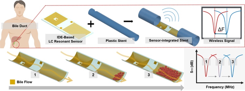
Schematic of the wireless biliary stent monitoring system and its resonance frequency response characteristics according to the degree of sludge accumulation.
where L is the inductance of the planar spiral inductor and C is the capacitance of the IDE structure. As dielectric materials are deposited on the IDE surface, the local dielectric environment around the electrodes changes, resulting in an increase in the effective permittivity. Consequently, the resonance frequency decreases as described in Eq. (1). Therefore, the occurrence of surface deposition can be indirectly detected by tracking shifts in the resonance frequency. Furthermore, the sensor operates wirelessly through inductive coupling with an external antenna, eliminating the need for an internal power source. This feature makes it particularly suitable for implantable biomedical applications where wired connections or battery integration are difficult to implement.
2.2 Sensor Design and Fabrication
The design and fabrication process of the proposed wireless IDE sensor are shown in Fig. 2. As illustrated in Fig. 2(a), the sensor features a multilayer structure consisting of a KMSF 1000 base layer, Cr/Au metal interconnects, a KMSF 1000 insulation layer, and electroplated Cr/Au IDE electrodes. The target resonance frequency range for biliary stent applications was set between 80 and 120 MHz by considering the dimensional constraints of the device and fabrication requirements.
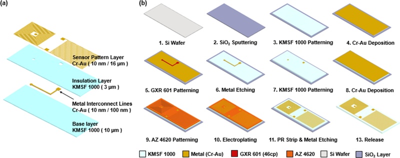
Design and fabrication process of the proposed wireless IDE sensor. (a) Schematic illustration of the sensor structure consisting of an IDE capacitor and a planar spiral inductor. (b) Fabrication process flow of the proposed IDE sensor.
According to previous studies, increasing the number of inductor turns leads to higher inductance and lower resonance frequencies, which in turn enhances the wireless recognition distance [16]. Therefore, the inductor in this study was designed to maximize the number of turns within the limited stent structure, while the IDE capacitance was adjusted to ensure operation within the target frequency range. The capacitance of the IDE structure was calculated using the analytical model proposed by Bahl [17], and the inductance was calculated using the model by Mohan [18]. The IDE electrodes were designed with 50 electrode pairs, an electrode length of 4,780 μm, an electrode width of 60 μm, and a spacing of 80 μm. For theoretical calculations, the effective permittivity was assumed to be 1. The inductor was designed with 20 turns, an outer diameter of 5,000 μm, an inner diameter of 980 μm, and a spacing of 40 μm.
Based on these analytical models, the capacitance and inductance were estimated to be 1.8681 pF and 1.2449 μH, respectively, resulting in a theoretical resonance frequency of approximately 104.37 MHz. Since the Bahl model is a two-dimensional approximation that does not account for electrode thickness, the actual capacitance of the fabricated IDE structure may differ slightly from the calculated value. However, this discrepancy is expected to remain well within the target range of 80–120 MHz.
The dimensions of the proposed sensor were determined by considering both the structural constraints of commercial biliary stents and the stability of wireless resonance characteristics. The maximum diameter of a commercial biliary stent is approximately 3.3 mm (10 Fr), corresponding to a circumference of about 10.4 mm. If a sensor were attached around the entire circumference, the spiral inductor could overlap with itself, causing mutual coupling that degrades the wireless resonance performance. Therefore, the sensor width was designed to be 5 mm so that it occupies approximately half of the stent circumference. The length of the sensor was determined to provide sufficient area for the number of IDE fingers and inductor turns required to achieve the necessary capacitance and inductance. This design ensures the target resonance frequency while maintaining stable wireless characteristics and sensor sensitivity.
The fabrication process is summarized in Fig. 2(b). The sensor was fabricated on a SiO₂-coated silicon wafer using MEMS processes. First, a KMSF 1000 base layer was formed, followed by the patterning of Cr/Au metal interconnects. A 3-μm-thick KMSF 1000 insulation layer was used to electrically isolate the IDE electrodes from the metal interconnects. To form the IDE electrodes, Au electroplating was performed using an AZ 4620 mold on a Cr/Au seed layer with an applied current of 0.08 A for 2 hours. Finally, the SiO₂ sacrificial layer was etched using buffered oxide etchant (BOE) to release the sensor from the wafer. The fabricated sensor is shown in Fig. 3, and the released device was attached to a mock plastic biliary stent with a diameter of 5 mm, as shown in Fig. 3(a). The morphology of the electroplated electrodes was confirmed via optical microscopy and cross-sectional SEM analysis, which verified an electrode thickness of approximately 16 μm (Fig. 3(b, c)).
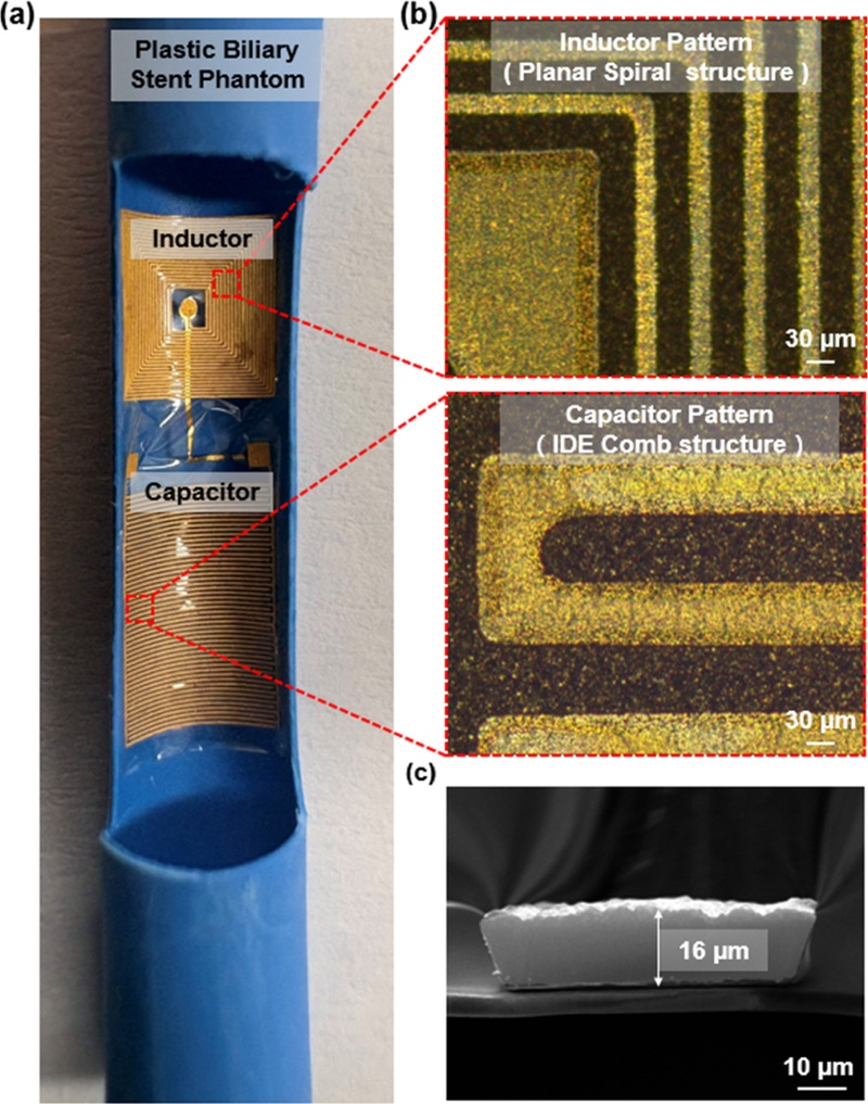
Fabrication results of the proposed wireless IDE sensor. (a) Fabricated sensor attached to a mock plastic biliary stent. (b) Optical microscope image of the electroplated IDE electrodes. (c) Cross-sectional SEM image showing the electrode thickness.
2.3 Experimental Setup
The performance of the fabricated sensor was evaluated using an external reader coil connected to a vector network analyzer (VNA, E5061B). The sensor was placed above the reader coil to measure resonance characteristics via inductive coupling. The resonance frequency was extracted from the S11 parameters to analyze the response to changes in dielectric properties around the IDE electrodes. Initially, measurements were performed in a flat configuration to verify the basic resonance characteristics. Subsequently, to mimic the actual application environment, the sensor was fixed to a mock plastic stent using epoxy adhesive, and its performance was evaluated under the same conditions.
Human bile is a complex biofluid predominantly composed of water (approximately 95%), with the remaining portion consisting of bile acids, cholesterol, and lipids [6,7,20]. Accordingly, DI water was first employed as a baseline to evaluate the fundamental dielectric sensitivity of the IDE sensor. To further account for the non-aqueous constituents, a water–lipid mixture was used to investigate the influence of hydrophobic residues, primarily cholesterol and fatty acids that persist on the sensor surface after fluid passage. Furthermore, clinical biliary sludge is characterized by a proteinaceous matrix where glycoproteins such as mucin act as a biological glue, facilitating the adherence of bacterial colonies and calcium-based crystals to the stent wall [7]. To model this organic framework, collagen was selected not to reproduce the exact biochemical composition of biliary sludge but to serve as a controllable surrogate material capable of forming uniform and repetitive deposition layers. This allows for a systematic investigation of the IDE sensor’s response to progressive structural accumulation and adherence of solid deposits. Nevertheless, these simplified surrogate materials have limitations in fully replicating the comprehensive biochemical complexity and dynamic biological variability of actual clinical bile.
The experiments first evaluated the response according to the water coverage area on the electrode surface. Then, the resonance characteristics under water-only and water–lipid mixture conditions were compared to investigate the influence of surface residues. Furthermore, collagen was employed as a model residue material to simulate progressive deposit accumulation. The accumulation layer was formed by repeating the deposition and drying of collagen solutions, and the resonance frequency shift was measured at each step. All experiments were conducted at room temperature under low-flow conditions using a precision micropipette to reflect the physiological characteristics of the biliary tract.
3. RESULTS AND DISCUSSIONS
3.1 Resonance Response According to Water Coverage Area
Fig. 4 shows the variation in the resonance frequency of the IDE sensor as the water coverage area on the sensor surface was gradually increased. To investigate the effect of the dielectric coverage range on the sensor response, the sensing region was divided into four identical sections, and water was sequentially applied to each section. As the water coverage area increased, the resonance frequency gradually decreased from approximately 87.35 MHz to 36.78 MHz, resulting in a total frequency shift of approximately 50.57 MHz. The most significant frequency shift was observed during the initial deposition stage, with the magnitude of the shift progressively decreasing as the covered area expanded.
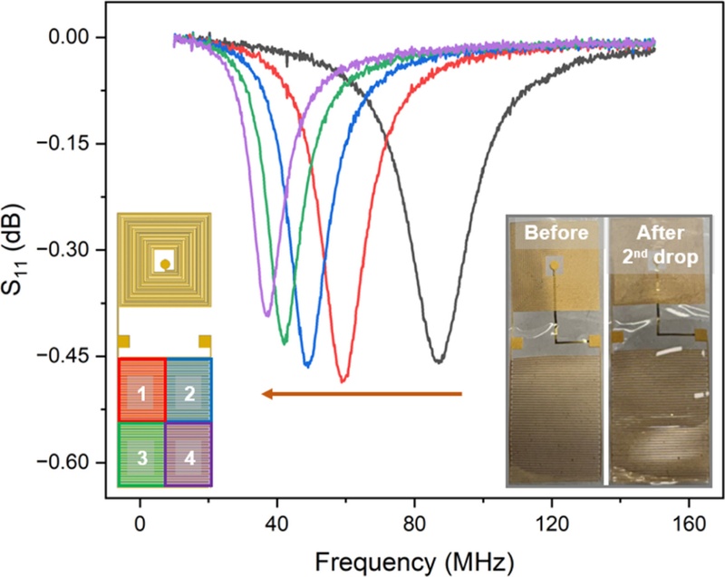
Resonance frequency variation of the IDE sensor according to the water coverage area on the electrode surface.
This phenomenon can be explained by the distribution of the fringing electric field in the IDE structure. When water first contacts the IDE electrodes, the high dielectric constant of water causes a rapid change in the local dielectric environment, leading to a substantial increase in capacitance. As the coverage area increases further, the effective dielectric environment approaches a saturation state, which reduces the rate of frequency shift. As described in Eq. (1), an increase in capacitance leads to a decrease in resonance frequency. Therefore, the observed frequency reduction confirms that the proposed IDE sensor effectively responds to permittivity changes occurring near the electrode surface.
3.2 Comparison of Resonance Characteristics Under Water and Water–Lipid Mixture Conditions
To evaluate the feasibility of the proposed sensor for biliary stent monitoring, resonance frequency responses were investigated under water-only and water–lipid mixture conditions, as shown in Figs. 5(a) and (b). Under the water-only condition in Fig. 5(a), the resonance frequency decreased immediately upon fluid exposure due to an increase in the effective permittivity around the IDE electrodes, with a maximum frequency shift of approximately 55.65 MHz. As the fluid supply was terminated and the water evaporated, the resonance frequency gradually recovered, returning to approximately 71.25 MHz—a value close to its initial state—after 10 minutes.
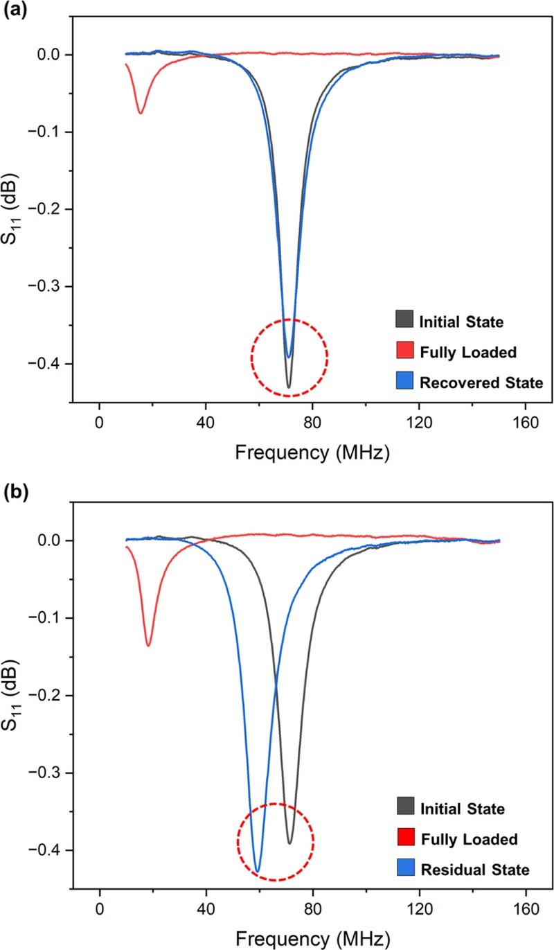
Resonance frequency responses of the IDE sensor under different fluid conditions. (a) Water-only condition. (b) Water–lipid mixture condition.
In contrast, the water–lipid mixture condition shown in Fig. 5(b) exhibited different recovery characteristics. Upon fluid application, the resonance frequency decreased by approximately 53.03 MHz, which is comparable to the water-only condition. However, even 10 minutes after fluid removal, the resonance frequency did not fully recover and remained shifted by approximately 11.9 MHz from the initial value. This incomplete recovery is attributed to the lipid components remaining on the IDE electrode surface after the liquid was removed. These residues altered the local dielectric environment around the electrodes, inducing a persistent change in capacitance. Consequently, the resonance frequency remained lower than its initial value even in the absence of the liquid medium.
These results indicate that the proposed IDE sensor can distinguish not only transient permittivity changes caused by liquid exposure but also residual surface deposits. Considering that biliary sludge consists of various organic components such as cholesterol, phospholipids, and mucin, these sensor characteristics are expected to be useful for detecting sludge accumulation during the early stages of biliary stent occlusion.
3.3 Resonance Response to Repeated Formation of Collagen-Based Model Fouling Layers
The resonance frequency response of the IDE sensor during the repeated formation of collagen-based model fouling layers is shown in Fig. 6. In this experiment, collagen was used as a model material to simulate a repeated surface deposition environment. First, approximately 0.2 mL of water was applied to establish the baseline resonance frequency. Subsequently, 0.2 mL of a 5 wt.% collagen solution was selectively applied to the capacitor region, held for 4 minutes, and then dried for 10 minutes to evaporate the solvent and form the model fouling layer. This process was repeated nine times, with the resonance frequency measured under the same water application conditions after every three cycles. The estimated total amount of collagen applied over nine deposition cycles was approximately 90 mg, resulting in a total frequency shift of 19.25 MHz. Based on the estimated applied amount, the frequency response normalized to the collagen amount was calculated to be approximately 0.214 MHz/mg. Because the actual amount of collagen retained on the sensor surface was not directly measured, this value represents the sensor response under the present experimental conditions rather than a clinically validated mass sensitivity. Moreover, quantitative data on the total amount of sludge accumulated in retrieved clinical biliary stents remain limited, preventing a reliable direct comparison with clinical sludge accumulation.
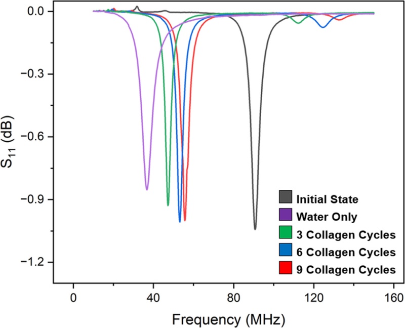
Resonance frequency response of the IDE sensor to repeated collagen deposition cycles.
The experimental results showed that before collagen treatment, water application induced a frequency shift of approximately 54.08 MHz. However, as collagen deposition was repeated, the frequency shift caused by water application gradually decreased. After nine deposition cycles, the frequency shift was reduced to approximately 34.83 MHz, representing a 35.6% decrease compared to the initial response. This reduction is attributed to changes in the local dielectric properties around the electrodes caused by the accumulated collagen layer. Prior to deposition, the large dielectric contrast between air and water resulted in a sharp increase in capacitance and a significant decrease in resonance frequency. However, as collagen accumulated on the sensor surface, the local dielectric environment gradually changed, which in turn reduced the additional variation in effective permittivity during subsequent water exposure. Consequently, as the amount of accumulated collagen increased, the resonance frequency response to the same volume of liquid application became progressively smaller.
These findings demonstrate that the proposed IDE sensor reacts sensitively not only to simple liquid exposure but also to the progressive accumulation of materials on the electrode surface. The ability to detect local dielectric changes caused by collagen-based model fouling layers supports the applicability of the IDE-based wireless sensor for monitoring surface deposition associated with sludge accumulation in biliary stents. Meanwhile, since actual biliary stents are used in the body for several weeks to months, further validation of the stability and reliability of the sensor in long-term environments is required. Although the proposed IDE sensor exhibited clear wireless resonance responses under the present laboratory conditions, its practical in vivo readout performance may vary because the sensor operates through inductive coupling with an external reader coil. An increased distance between the reader coil and the implanted sensor, as well as angular or positional misalignment between the two coils, may reduce the coupling efficiency and weaken the measured resonance response. In addition, surrounding biological tissues may alter the electromagnetic coupling environment and attenuate the received signal. Variations in the relative position of the reader coil and the sensor during repeated measurements may also affect the stability of the measured S11 response. Therefore, further optimization of the reader-coil geometry, readout distance, alignment tolerance, and measurement configuration will be required to achieve stable wireless interrogation under in vivo conditions.
4. CONCLUSIONS
In this study, an IDE-based LC resonant sensor was applied to monitor surface deposition on biliary stents, and the resonance responses were analyzed using water, water–lipid mixtures, and collagen-based model fouling layers. The proposed sensor was fabricated using MEMS processes and designed with a planar spiral inductor and an IDE capacitor connected in series to enable passive wireless sensing through resonance frequency measurements.
Initially, an investigation of the response according to the water coverage area on the IDE surface confirmed that the resonance frequency decreased progressively as the covered area increased. This demonstrates that the sensor is highly sensitive to permittivity changes around the electrodes. Furthermore, experiments under water-only and water–lipid mixture conditions showed that the sensor could effectively detect both transient dielectric changes during fluid exposure and residual materials remaining on the surface after fluid removal.
In the repeated collagen deposition experiments conducted to evaluate the response to progressive material accumulation, the frequency shift in response to water exposure gradually decreased as the amount of collagen increased. This indicates that the sensor can precisely detect the local dielectric environment altered by surface deposits.
These results confirm the feasibility of using wireless IDE sensors for monitoring the internal conditions of biliary stents. This work provides a foundation for developing implantable wireless monitoring systems for the early detection of deposit accumulation associated with biliary stent occlusion. Future studies will focus on evaluating sensor performance under conditions reflecting actual biliary flow rates and further validating the effectiveness of the sensing mechanism in environments where actual sludge is formed. In addition, long-term evaluations under prolonged and repeated fouling conditions will be required to further assess the stability and reproducibility of the proposed sensing approach.
Acknowledgments
This work was supported by the National Research Foundation of Korea (NRF) grant funded by the Korean government (MSIT) (RS-2022-NR072456).
REFERENCES
-
K. Yang, W. Sun, L. Cui, Y. Zou, C. Wen, R. Zeng, Advances in functional coatings on biliary stents, Regen. Biomater. 11 (2024) rbae001.
[https://doi.org/10.1093/rb/rbae001]

-
D. Ni, Q. Yang, L. Nie, J. Xu, S. He, J. Yao, The past, present, and future of endoscopic management for biliary strictures: technological innovations and stent advancements, Front. Med. 11 (2024) 1334154.
[https://doi.org/10.3389/fmed.2024.1334154]
-
European Association for the Study of the Liver, EASL Clinical Practice Guidelines: management of cholestatic liver diseases, J. Hepatol. 51 (2009) 237–267.
[https://doi.org/10.1016/j.jhep.2009.04.009]
-
A. Singh, A. Gelrud, B. Agarwal, Biliary strictures: diagnostic considerations and approach, Gastroenterol. Rep. 3 (2015) 22–31.
[https://doi.org/10.1093/gastro/gou072]
-
S.Y. Lee, S.I. Jang, M.J. Chung, J.H. Cho, M.Y. Do, H.S. Lee, et al., A short fully covered self-expandable metal stent for management of benign biliary stricture not caused by living-donor liver transplantation, J. Clin. Med. 13 (2024) 1186.
[https://doi.org/10.3390/jcm13051186]
-
G. Donelli, E. Guaglianone, R. Di Rosa, F. Fiocca, A. Basoli, Plastic biliary stent occlusion: factors involved and possible preventive approaches, Clin. Med. Res. 5 (2007) 53–60.
[https://doi.org/10.3121/cmr.2007.683]
-
J. Gruszecka, R. Filip, Bacterial biofilms—a threat to biliary stents, understanding their formation, clinical consequences and management, Medicina 61 (2025) 512.
[https://doi.org/10.3390/medicina61030512]
-
J.J. Bergman, E.A. Rauws, J.G. Tijssen, G.N. Tytgat, K. Huibregtse, Biliary endoprostheses in elderly patients with endoscopically irretrievable common bile duct stones: report on 117 patients, Gastrointest. Endosc. 42 (1995) 195–201.
[https://doi.org/10.1016/S0016-5107(95)70091-9]
-
G. Costamagna, M. Pandolfi, M. Mutignani, C. Spada, V. Perri, Long-term results of endoscopic management of postoperative bile duct strictures with increasing numbers of stents, Gastrointest. Endosc. 54 (2001) 162–168.
[https://doi.org/10.1016/S0016-5107(01)70100-9]
-
X.B. Cai, W.X. Zhang, X.J. Wan, Q. Yang, X.S. Qi, X.P. Wang, et al., The effect of a novel drug-eluting plastic stent on biliary stone dissolution in an ex vivo bile perfusion model, Gastrointest. Endosc. 79 (2014) 156–162.
[https://doi.org/10.1016/j.gie.2013.09.013]
-
M. Sekine, M. Ijima, S. Noguchi, E. Kurihara, T. Kobatake, T. Mizutani, et al., Efficacy of a novel dual-layer plastic stents for malignant biliary obstruction, J. Clin. Med. 14 (2025) 764.
[https://doi.org/10.3390/jcm14030764]
-
R.C. Ooi, X.Y. Luo, S.B. Chin, A.G. Johnson, N.C. Bird, The flow of bile in the human cystic duct, J. Biomech. 37 (2004) 1913–1922.
[https://doi.org/10.1016/j.jbiomech.2004.02.029]
-
A. Esteller, Physiology of bile secretion, World J. Gastroenterol. 14 (2008) 5641–5649.
[https://doi.org/10.3748/wjg.14.5641]
-
J.L. Boyer, C.J. Soroka, Bile formation and secretion: An update, J. Hepatol. 75 (2021) 190–201.
[https://doi.org/10.1016/j.jhep.2021.02.011]
-
R.M. Nambisan, S.R. Green, R.S. Kwon, G.H. Elta, Y.B. Gianchandani, A microsystem for in vivo wireless monitoring of plastic biliary stents using magnetoelastic sensors, Microsyst. Nanoeng. 10 (2024) 159.
[https://doi.org/10.1038/s41378-024-00772-8]
-
N.E. Oyunbaatar, D.S. Kim, G. Prasad, Y.J. Jeong, D.W. Lee, Self-rollable polymer stent integrated with wireless pressure sensor for real-time monitoring of cardiovascular pressure, Sens. Actuators A Phys. 346 (2022) 113869.
[https://doi.org/10.1016/j.sna.2022.113869]
- I.J. Bahl, Lumped Elements for RF and Microwave Circuits, 2nd ed., Artech House, Norwood, MA, 2022.
-
S.S. Mohan, M. del Mar Hershenson, S.P. Boyd, T.H. Lee, Simple accurate expressions for planar spiral inductances, IEEE J. Solid-State Circuits 34 (1999) 1419–1424.
[https://doi.org/10.1109/4.792620]
-
R. Igreja, C.J. Dias, Analytical evaluation of the interdigital electrodes capacitance for a multi-layered structure, Sens. Actuators A Phys. 112 (2004) 291–301.
[https://doi.org/10.1016/j.sna.2004.01.040]
-
B. Sanjel, W.S. Shim, Recent advances in understanding the molecular mechanisms of cholestatic pruritus: A review, Biochim. Biophys. Acta Mol. Basis Dis. 1866 (2020) 165958.
[https://doi.org/10.1016/j.bbadis.2020.165958]