
Recent Advances in AI-Integrated SERS Sensors for Biomedical Diagnostics

ⓒ The Korean Sensors Society
This is an Open Access article distributed under the terms of the Creative Commons Attribution Non-Commercial License (https://creativecommons.org/licenses/by-nc/3.0/) which permits unrestricted non-commercial use, distribution, and reproduction in any medium, provided the original work is properly cited.
Abstract
Surface-enhanced Raman scattering (SERS) has emerged as a promising sensing technology for biomedical diagnostics owing to its ability to provide ultrasensitive molecular fingerprint information from complex biological samples. However, conventional SERS-based biosensors still face limitations such as spectral complexity, signal variability, and expert-dependent interpretation. Recently, the integration of artificial intelligence (AI) with SERS platforms has attracted increasing attention for automated and accurate spectral analysis. By combining machine learning algorithms with spectral preprocessing techniques, AI-assisted SERS (AI-SERS) sensors enable rapid and reliable classification of biological samples. This semi-review summarizes recent advances in AI-integrated SERS sensors for biomedical applications, including cancer diagnostics, pathogen detection, and biofluid analysis. Particular emphasis is placed on cerebrospinal fluid (CSF) rhinorrhea diagnosis, where AI-SERS platforms can effectively distinguish CSF from nasal secretions despite their highly similar biochemical characteristics. In addition, portable Raman systems and cross-instrument spectral analysis are discussed for future point-of-care diagnostic applications.
Keywords:
Surface-enhanced Raman scattering, Artificial intelligence, Biosensor, Cerebrospinal fluid rhinorrhea1. INTRODUCTION
Raman spectroscopy has attracted significant attention in the biomedical sensing field owing to its ability to provide molecular fingerprint information through non-destructive and label-free analysis [1,2]. Since Raman signals originate from the intrinsic vibrational modes of molecules, Raman spectroscopy enables highly selective chemical identification of complex biological samples. However, the inherently weak Raman scattering efficiency often limits its practical sensitivity for trace-level biomarker detection and clinical diagnostics [3,4]. To overcome this limitation, surface-enhanced Raman scattering (SERS) has been extensively investigated as an advanced spectroscopic sensing platform capable of dramatically amplifying Raman signals through localized surface plasmon resonance generated on noble metal nanostructures such as gold (Au) and silver (Ag) [5].
Owing to its ultrasensitive detection capability, SERS has been widely applied to various biomedical applications, including cancer diagnostics, pathogen detection, and biofluid analysis [6-8]. Recent advances in nanofabrication technologies have enabled the development of high-performance plasmonic substrates with improved signal enhancement and reproducibility [5,9]. Nevertheless, conventional SERS-based biosensors still face several limitations that hinder their practical clinical translation. Biological samples typically exhibit highly complex and overlapping spectral features, making accurate interpretation challenging. In addition, signal variability caused by substrate heterogeneity, environmental conditions, and instrument-dependent spectral differences often reduces analytical reliability [10,11]. Consequently, conventional spectral interpretation frequently relies on expert-dependent analysis, limiting rapid and automated diagnosis.
Recently, artificial intelligence (AI)-assisted spectral analysis has emerged as a promising strategy for overcoming the limitations of conventional SERS sensing systems. Machine learning and deep learning algorithms can efficiently extract hidden spectral patterns from complex Raman datasets and enable automated classification of biological samples with high accuracy [12,13]. By integrating AI with SERS platforms, intelligent sensing systems capable of rapid, reproducible, and user-independent diagnosis have become increasingly feasible. Furthermore, recent studies have demonstrated the potential of portable Raman devices combined with AI-driven spectral processing for point-of-care biomedical applications [14-16].
Among emerging biomedical applications, cerebrospinal fluid (CSF) rhinorrhea diagnosis has recently attracted attention as a representative target for AI-SERS sensing. CSF rhinorrhea is characterized by leakage of cerebrospinal fluid through the nasal cavity and is often difficult to distinguish from normal nasal secretions because of their visually similar characteristics [17,18]. Delayed diagnosis can increase the risk of severe complications such as meningitis due to pathogen penetration into the central nervous system [19]. Therefore, rapid and accurate discrimination between CSF and nasal secretions is clinically important. Recent studies have demonstrated that AI-integrated SERS platforms can effectively classify these highly similar biofluids through intelligent spectral analysis [20].
In this review, recent advances in AI-integrated SERS sensors for biomedical diagnostics are summarized, with particular emphasis on machine learning-assisted spectral analysis and plasmonic substrate engineering strategies. In particular, cerebrospinal fluid (CSF) rhinorrhea is highlighted in this review as a representative case study because it involves the discrimination of two visually similar and biochemically overlapping biofluids, CSF and nasal secretion. This diagnostic challenge clearly illustrates the strength of AI-SERS sensing, which can extract subtle molecular differences from complex spectra and provide rapid classification beyond conventional visual or peak-based interpretation.
2. CORE COMPONENTS OF AI-SERS SENSORS
2.1 Principle of Surface-Enhanced Raman Scattering (SERS)
Surface-enhanced Raman scattering (SERS) is an advanced spectroscopic sensing technique capable of significantly amplifying Raman signals through plasmonic interactions occurring on metallic nanostructures [21]. As shown in Fig. 1(a), Raman signals inherently have low scattering efficiency, which limits their direct use as a sensing technology. To overcome this, SERS amplifies the local electromagnetic field near nanostructured metal surfaces, particularly surfaces composed of gold (Au) and silver (Ag), enabling high-sensitivity molecular detection. When incident light excites localized surface plasmon resonance (LSPR) on metallic nanostructures, strong electromagnetic fields are generated in nanoscale junctions, commonly referred to as “hotspots,” where Raman signal enhancement can increase by several orders of magnitude [22].
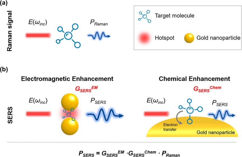
Schematic diagram of the SERS mechanism. Difference between (a) Raman signal and (b) SERS signal is highlighted with electromagnetic and chemical enhancement.
The signal enhancement mechanism of SERS is generally explained by two major contributions: electromagnetic enhancement and chemical enhancement (Fig. 1(b)). Electromagnetic enhancement primarily originates from localized plasmonic field amplification around metallic nanostructures and is considered the dominant mechanism responsible for high signal enhancement. Chemical enhancement, although relatively smaller in magnitude, arises from charge-transfer interactions between target molecules and metal surfaces, contributing to improved Raman scattering efficiency. In biomedical sensing applications, the synergistic effects of these mechanisms enable ultrasensitive detection of biomolecules even at trace concentrations [1].
To maximize sensing performance, considerable efforts have been devoted to engineering plasmonic substrates capable of generating dense and reproducible hotspots [23]. Various nanostructures, including nanoparticles, nanopillars, nanogaps, and hierarchical hybrid architectures, have been developed to optimize plasmonic coupling and signal reproducibility. In particular, Au and Ag-based hybrid nanostructures have attracted increasing attention owing to their ability to combine strong plasmonic enhancement with improved structural stability, thereby facilitating reliable sensing performance in complex biological environments [24].
2.2 Artificial Intelligence-Assisted Spectral Analysis
Despite the remarkable sensitivity of SERS, the interpretation of Raman spectra from biological samples remains challenging because biological fluids often contain highly overlapping molecular fingerprints and complex biochemical information [25,26]. Conventional spectral analysis typically depends on peak assignment and expert interpretation, which may limit rapid and reproducible diagnosis [25]. Moreover, variations caused by sample heterogeneity, environmental conditions, and instrumental differences further complicate reliable classification.
To overcome these limitations, artificial intelligence (AI)-assisted spectral analysis has emerged as a powerful strategy for extracting hidden information from complex Raman datasets [3,16]. Machine learning algorithms can automatically identify subtle spectral differences that may not be easily distinguishable through conventional approaches. Classical machine learning methods, such as principal component analysis (PCA), support vector machines (SVM), random forest (RF), and logistic regression (LR), have been widely employed for spectral classification and biomarker discrimination [27,28]. More recently, deep learning approaches, including convolutional neural networks (CNNs), have demonstrated promising performance in automated feature extraction and classification without extensive manual intervention. Among these approaches, machine learning-assisted Raman analysis offers particular advantages for biomedical sensing because it enables rapid, objective, and user-independent decision-making. By learning hidden spectral relationships from large datasets, AI-based algorithms can significantly improve diagnostic accuracy and robustness, supporting the development of intelligent biosensing systems [29].
2.3 Spectral Preprocessing for AI-SERS Sensors
Reliable AI-SERS sensing requires not only robust machine learning algorithms but also appropriate preprocessing of Raman spectra. Raw Raman data often contain fluorescence background, noise, baseline drift, and intensity variation caused by experimental conditions or instrument settings. Without suitable preprocessing, these unwanted spectral variations may reduce model performance and classification reliability.
Accordingly, preprocessing techniques such as baseline correction, smoothing, normalization, denoising, and dimensionality reduction are commonly applied before machine learning analysis [30]. As shown in Fig. 2, baseline correction is frequently used to remove fluorescence interference, whereas normalization techniques help minimize intensity fluctuations among measurements. In addition, dimensionality reduction approaches such as PCA can simplify complex spectral datasets and facilitate efficient classification.
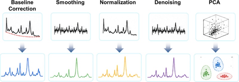
Schematic representation of representative spectral preprocessing procedures for AI-SERS analysis, including baseline correction, smoothing, normalization, denoising, and dimensionality reduction. These preprocessing strategies improve spectral quality and enhance feature extraction for reliable machine learning-assisted classification of Raman signals.
Another emerging challenge in AI-SERS sensing involves instrument-dependent variability, including differences in spectral range, resolution, and acquisition conditions among Raman systems. Such inconsistencies may hinder model transferability and limit practical deployment across different sensing platforms. Therefore, recent studies have increasingly focused on cross-instrument spectral standardization strategies to improve robustness and enable portable point-of-care Raman diagnostics [31,32]. By combining optimized preprocessing with AI-assisted analysis, AI-SERS sensors are evolving into reliable and intelligent platforms for biomedical sensing applications.
3. BIOMEDICAL APPLICATIONS OF AI-SERS SENSORS
Artificial intelligence-assisted surface-enhanced Raman scattering (AI-SERS) sensors have recently emerged as powerful platforms for biomedical diagnostics by combining ultrasensitive molecular fingerprinting with automated spectral interpretation. In contrast to conventional Raman analysis, which often requires expert interpretation and suffers from complex spectral overlap, AI-assisted approaches enable rapid classification of biological samples through machine learning-driven pattern recognition [33]. Owing to these advantages, AI-SERS sensors have been increasingly investigated for cancer diagnosis, pathogen detection, and biofluid-based disease screening.
3.1 Cancer Diagnostics
Cancer diagnosis represents one of the most actively explored biomedical applications of AI-SERS sensing. Because tumor progression is accompanied by subtle biochemical alterations in serum, plasma, extracellular vesicles, and other biological matrices, SERS-based molecular fingerprinting has shown considerable potential for non-invasive cancer screening. However, cancer-associated spectral variations are often weak and highly overlapped, limiting reliable interpretation through conventional peak-based analysis. Machine learning algorithms have therefore been widely integrated with SERS to extract hidden spectral information and improve diagnostic accuracy [34].
For example, a deep learning-assisted SERS platform demonstrated accurate multi-cancer detection using plasma-derived exosomes and AI-based spectral analysis [35]. The proposed framework enabled simultaneous classification of multiple cancer types and tissue-of-origin prediction using label-free exosomal SERS fingerprints, demonstrating the feasibility of intelligent liquid biopsy systems for early cancer diagnosis.
Similarly, serum-based AI-SERS approaches have been reported for lung cancer screening using convolutional neural networks (CNNs) and multivariate classification strategies [36]. These systems achieved high diagnostic performance by learning disease-specific spectral signatures from complex serum samples and demonstrated the potential of AI-SERS platforms for postoperative monitoring and recurrence prediction.
3.2 Pathogen and Infectious Disease Detection
AI-SERS sensors have also attracted considerable attention for rapid pathogen identification and infectious disease diagnosis. Traditional microbiological assays generally require time-consuming culturing procedures or expensive molecular analysis, whereas AI-SERS systems enable rapid, label-free classification of pathogens based on molecular vibrational fingerprints [37]. Since microbial Raman spectra frequently exhibit subtle interspecies differences and strong spectral overlaps, machine learning-based analysis is particularly valuable for improving diagnostic reliability.
Recent studies have demonstrated the feasibility of AI-assisted SERS platforms for viral classification, including respiratory viruses and coronavirus detection [38]. For example, convolutional neural network (CNN)-based Raman classification enabled accurate differentiation of viral species and subtypes using subtle spectral differences associated with viral proteins and lipid structures. Likewise, machine learning-assisted SERS strategies have been applied to SARS-CoV-2 screening, supporting rapid and automated infectious disease diagnosis.
3.3 Biofluid-Based Diagnostics
Beyond cancer and pathogen detection, AI-SERS sensing has been increasingly explored for biofluid-based diagnostics because many diseases induce biochemical changes that can be reflected in blood, urine, saliva, tears, and nasal secretions [39]. Since Raman spectroscopy is minimally affected by water interference and requires limited sample preparation, SERS-based biofluid analysis provides a promising route toward non-invasive and rapid diagnosis.

Representative studies of biomedical AI-SERS sensors.
In particular, urine- and serum-based AI-SERS systems have demonstrated promising performance for disease screening through multivariate statistical analysis and machine learning classification [40]. Biofluid-derived molecular fingerprints combined with AI analysis allow discrimination between healthy and disease states while reducing user dependency and diagnostic subjectivity. More importantly, recent progress in portable Raman instrumentation suggests the possibility of translating AI-SERS sensing into point-of-care diagnostic systems.
Overall, recent studies suggest that AI-SERS sensors can significantly improve the interpretability and diagnostic performance of conventional Raman biosensing systems by enabling automated feature extraction and intelligent classification. While current applications are primarily focused on cancer screening, pathogen identification, and biofluid analysis, the continued advancement of portable Raman systems and cross-platform AI processing is expected to further accelerate clinical translation. In this context, CSF rhinorrhea diagnosis represents an emerging example in which AI-SERS sensing can effectively distinguish highly similar biofluids for rapid clinical decision-making.
4. AI-SERS SENSOR FOR CEREBROSPINAL FLUID RHINORRHEA DIAGNOSIS
In this section, CSF rhinorrhea diagnosis is discussed not merely as an individual application but as a representative example demonstrating the practical value of AI-SERS sensing for distinguishing highly similar biological matrices. This case is particularly relevant because the diagnostic problem requires rapid, minimally invasive, and objective discrimination between CSF leakage and normal nasal secretion, which directly matches the core advantages of AI-integrated SERS platforms.
4.1 Clinical Need for Rapid CSF Rhinorrhea Screening
CSF rhinorrhea is a pathological condition characterized by leakage of cerebrospinal fluid into the nasal cavity through skull base defects. Because leaked CSF commonly appears as transparent nasal discharge, differentiation from benign nasal secretions using visual inspection alone is often difficult. Delayed diagnosis may lead to severe complications, including bacterial meningitis and intracranial infection, due to the direct communication established between the external environment and the central nervous system [41]. Conventional diagnostic strategies, including biochemical assays and imaging modalities, frequently require invasive procedures, extended examination time, or specialized infrastructure, limiting their accessibility in rapid clinical screening settings.
Given these challenges, there has been growing interest in rapid molecular sensing approaches capable of distinguishing CSF from nasal secretions in a minimally invasive manner. Raman spectroscopy and SERS-based analysis offer a promising alternative because they provide label-free molecular fingerprint information without requiring extensive sample preparation [42,43]. However, owing to the substantial biochemical similarity between CSF and nasal secretions, direct spectral discrimination remains challenging, thereby motivating the incorporation of AI-assisted analytical frameworks.
4.2 AI-SERS Strategy for Biofluid Discrimination
Recent studies have demonstrated that integrating SERS with machine learning enables discrimination of highly similar biofluids through the recognition of subtle spectral variations that are difficult to resolve by conventional inspection [20]. In the context of CSF rhinorrhea, an AI-SERS sensing workflow has been proposed in which patient-derived samples are directly deposited onto plasmonic SERS substrates, followed by Raman acquisition and automated spectral classification. Rather than relying on a single diagnostic biomarker, the approach interprets collective spectral patterns generated from multiple biochemical constituents present in biological fluids, including proteins, glycoproteins, lipids, and nucleic acid-related components.
As a representative strategy, signal stability was enhanced under physiologically suitable conditions using a corrosion-resistant Au@Ag bimetallic nanopillar substrate, as shown in Fig. 3(a). Compared with conventional metallic SERS structures, hybrid plasmonic architectures may provide improved robustness and stronger signal amplification through synergistic electromagnetic coupling. Importantly, this platform enabled reproducible spectral acquisition from clinical CSF and nasal secretion specimens while maintaining compatibility with portable Raman systems.
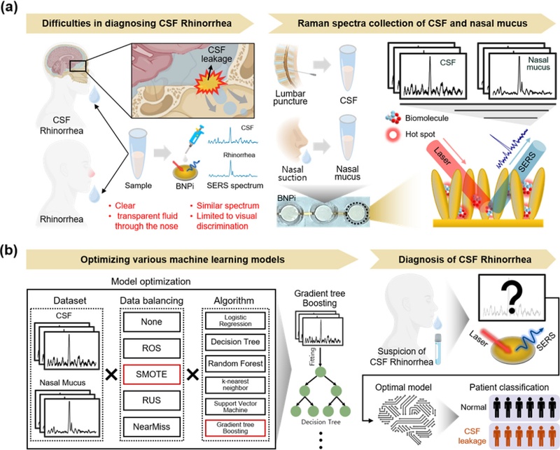
Schematic illustration of the AI-integrated SERS platform for cerebrospinal fluid (CSF) rhinorrhea diagnosis. (a) SERS-based molecular fingerprint acquisition using an Au@Ag nanopillar substrate. (b) AI-assisted spectral preprocessing and machine learning-based classification of CSF and nasal secretions.
4.3 Role of Artificial Intelligence in Spectral Classification
Although SERS provides rich biochemical information, direct comparison between CSF and nasal secretion spectra remains difficult because the overall spectrum exhibits substantial overlap. Several Raman bands associated with proteins, carbohydrates, lipids, and nucleic acid-related compounds may appear in both fluids with only modest intensity variation. Consequently, conventional peak-by-peak interpretation alone may be insufficient for reliable discrimination.
Artificial intelligence addresses this challenge by identifying subtle multidimensional spectral relationships beyond human visual interpretation (fig. 3(b)). Machine learning algorithms trained on SERS datasets can recognize hidden differences among highly correlated spectral features and transform these complex datasets into probabilistic diagnostic outputs. In representative AI-SERS studies for CSF rhinorrhea, supervised classification models demonstrated strong diagnostic performance during both internal and external validation, supporting the feasibility of intelligent Raman-assisted diagnosis in real clinical scenarios. Rather than depending on isolated spectral markers, diagnostic decisions were generated from integrated molecular signatures distributed across multiple Raman bands.
4.4 Toward Point-of-Care AI-SERS Diagnosis
One of the major advantages of AI-SERS sensing for CSF rhinorrhea diagnosis lies in its compatibility with portable diagnostic systems. As Raman instrumentation continues to evolve toward miniaturized and portable formats, recent studies have explored cross-instrument spectral standardization strategies to compensate for discrepancies in spectral range and resolution between benchtop and portable Raman devices. As illustrated in Fig. 4(a), although the same target analyte (4-aminothiophenol, 4-ATP) measured using benchtop and portable Raman systems exhibits identical characteristic Raman peaks, noticeable differences remain in spectral range and the number of data points corresponding to individual Raman bands. Such inconsistencies may reduce model transferability and limit the direct applicability of machine learning-assisted spectral classification across different devices.
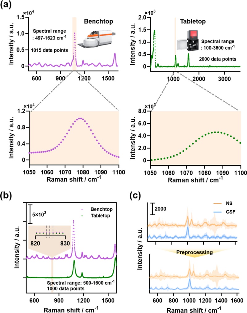
Description of the CISP (Cross-Instrument Spectrum Preprocessing) algorithm and platform validation using a portable Raman spectrometer. (a) SERS spectra and spectral resolution of 4-ATP measured with benchtop and portable Raman spectrometers. (b) Effect of CISP on SERS spectra of 4-ATP measured with different instruments. (c) SERS spectra of pre-treated cerebrospinal fluid (CSF) and physiological saline (NS).
To address these challenges, preprocessing and AI-based calibration strategies have been introduced to standardize instrument-dependent spectral variation, as shown in Fig. 4(b). By harmonizing spectral resolution, aligning data point density, and minimizing inter-device variability, these correction algorithms substantially improve the suitability of Raman spectra as training datasets for machine learning models. When applied to CSF and nasal secretion samples, the proposed framework reduced spectral variability and enhanced classification robustness, thereby facilitating more reliable discrimination between highly similar biological matrices (Fig. 4(c)).
Collectively, these findings highlight CSF rhinorrhea diagnosis as a representative example demonstrating how AI-assisted SERS sensing can bridge ultrasensitive molecular analysis with practical clinical translation. More broadly, this case underscores the increasingly important role of intelligent biosensing systems in addressing diagnostic challenges involving highly similar biological matrices that are difficult to differentiate using conventional analytical approaches.
5. CONCLUSIONS
The integration of AI with SERS has opened new opportunities for intelligent biomedical sensing by overcoming several longstanding limitations of conventional Raman analysis, including spectral complexity, signal variability, and user-dependent interpretation. By coupling ultrasensitive molecular fingerprint acquisition with automated data-driven classification, AI-SERS platforms enable rapid, objective, and highly reliable analysis of complex biological samples. As discussed throughout this semi-review, recent advances in plasmonic substrate engineering, spectral preprocessing strategies, and machine learning-assisted analysis have significantly improved the robustness and diagnostic capability of SERS-based sensing systems.
Particularly, biomedical applications involving cancer diagnosis, pathogen detection, and biofluid-based analysis highlight the growing utility of AI-SERS sensors for non-invasive and label-free diagnostics. Among these, CSF rhinorrhea diagnosis represents a compelling example demonstrating how AI-assisted Raman analysis can successfully distinguish highly similar biological matrices that remain difficult to classify through conventional analytical approaches. The integration of compelling Raman instrumentation and cross-platform spectral standardization further underscores the translational potential of AI-SERS sensing toward real-world clinical implementation.
Despite these advances, several challenges remain before widespread clinical adoption becomes feasible. The reproducibility and large-scale manufacturability of SERS substrates, standardization of spectral acquisition across instruments, availability of sufficiently diverse clinical datasets, and interpretability of AI-driven decisions continue to require further investigation. In particular, future research should move beyond isolated proof-of-concept demonstrations toward clinically robust sensing frameworks capable of maintaining diagnostic reliability across heterogeneous populations and measurement environments.
Looking forward, AI-SERS sensors are expected to evolve into next-generation intelligent biomedical sensing platforms capable of real-time, portable, and personalized diagnostics. Future systems may integrate miniaturized Raman hardware, adaptive machine learning models, cloud-based data analysis, and multimodal biosensing strategies to enable continuous health monitoring and point-of-care clinical decision support. Rather than functioning solely as analytical devices, AI-SERS technologies are likely to become intelligent diagnostic ecosystems that autonomously interpret molecular information and assist precision medicine. In this context, the convergence of AI and SERS is anticipated to play an increasingly important role in shaping future biomedical sensing technologies and accelerating the transition toward smarter and more accessible healthcare systems.
Acknowledgments
This research was funded by the National Research Foundation of Korea (NRF) under Grant Nos. RS-2023-NR077066, RS-2024-00438542 and RS-2025-00554830.
REFERENCES
-
R. Beeram, K.R. Vepa, V.R. Soma, Recent trends in SERS-based plasmonic sensors for disease diagnostics, biomolecules detection, and machine learning techniques, Biosensors 13 (2023) 328.
[https://doi.org/10.3390/bios13030328]

-
R. Pilot, R. Signorini, C. Durante, L. Orian, M. Bhamidipati, L. Fabris, A review on surface-enhanced Raman scattering, Biosensors 9 (2019) 57.
[https://doi.org/10.3390/bios9020057]
-
Y. Liu, Y. Wu, J. Wang, J. Qi, C. Zhou, Y. Xue, Recent Advances in Raman Spectral Classification with Machine Learning, Sensors 26 (2026) 341.
[https://doi.org/10.3390/s26010341]
-
J. Luo, J. Sun, H. Chen, C. Zhao, M. Ye, A Review of High-Sensitivity SERS-Active Photonic Crystal Fiber Sensors for Chemical and Biological Detection, Sensors 25 (2025) 6982.
[https://doi.org/10.3390/s25226982]
-
L.L. Lin, R. Alvarez-Puebla, L.M. Liz-Marzan, M. Trau, J. Wang, L. Fabris, et al., Surface-enhanced Raman spectroscopy for biomedical applications: recent advances and future challenges, ACS Appl. Mater. Interfaces 17 (2025) 16287–16379.
[https://doi.org/10.1021/acsami.4c17502]
-
K. Yuan, B. Jurado-Sánchez, A. Escarpa, Nanomaterials meet surface-enhanced Raman scattering towards enhanced clinical diagnosis: a review, J. Nanobiotechnology 20 (2022) 537.
[https://doi.org/10.1186/s12951-022-01711-3]
-
M. Constantinou, K. Hadjigeorgiou, S. Abalde-Cela, C. Andreou, Label-free sensing with metal nanostructure-based surface-enhanced Raman spectroscopy for cancer diagnosis, ACS Appl. Nano Mater. 5 (2022) 12276–12299.
[https://doi.org/10.1021/acsanm.2c02392]
-
N. Lyu, A. Hassanzadeh-Barforoushi, L.M. Rey Gomez, W. Zhang, Y. Wang, SERS biosensors for liquid biopsy towards cancer diagnosis by detection of various circulating biomarkers: current progress and perspectives, Nano Converg. 11 (2024) 22.
[https://doi.org/10.1186/s40580-024-00428-3]
-
K. Milenko, F.T. Dullo, P.C. Thrane, Z. Skokic, C.A. Dirdal, UV-nanoimprint lithography for predefined SERS nanopatterns which are reproducible at low cost and high throughput, Nanomaterials 13 (2023) 1598.
[https://doi.org/10.3390/nano13101598]
-
I.A. Bratchenko, L.A. Bratchenko, Overestimation of the classification model for Raman spectroscopy data of biological samples, Msystems 10 (2025) e00063–25.
[https://doi.org/10.1128/msystems.00063-25]
-
S. Lee, R. Park, H.S. Jung, AI‐enhanced surface‐enhanced raman scattering for accurate and sensitive biomedical sensing, Adv. Intell. Disc. 2 (2026) e202500030.
[https://doi.org/10.1002/aidi.202500030]
-
A. Lebrun, H. Fortin, N. Fontaine, D. Fillion, O. Barbier, D. Boudreau, Pushing the limits of surface-enhanced Raman spectroscopy (SERS) with deep learning: identification of multiple species with closely related molecular structures, Appl. Spectrosc. 76 (2022) 609–619.
[https://doi.org/10.1177/00037028221077119]
-
F. Ebrahimi, A. Kumari, K. Dellinger, Integration of nanoengineering with artificial intelligence and machine learning in surface‐enhanced Raman spectroscopy (SERS) for the development of advanced biosensing platforms, Adv. Sensor Res. 4 (2025) 2400155.
[https://doi.org/10.1002/adsr.202400155]
-
C. Soliman, D. Tu, S. Mabbott, G. Coté, K. Maitland, Portable, multi-modal Raman and fluorescence spectroscopic platform for point-of-care applications, J. Biomed. Opt. 27 (2022) 095006.
[https://doi.org/10.1117/1.JBO.27.9.095006]
-
M. Fan, Y. Weng, Y. Liu, Y. Lu, L. Xu, J. Ye, et al., Surface‐Enhanced Raman Spectroscopy‐Based Optical Biosensor for Liquid Biopsy: Toward Precision Medicine, Laser Photonics Rev. 18 (2024) 2301072.
[https://doi.org/10.1002/lpor.202301072]
-
T.R. Szymborski, S.M. Berus, A.B. Nowicka, G. Słowiński, A. Kamińska, Machine learning for COVID-19 determination using surface-enhanced raman spectroscopy, Biomedicines 12 (2024) 167.
[https://doi.org/10.3390/biomedicines12010167]
- A. Abuabara, Cerebrospinal fluid rhinorrhoea: diagnosis and management, Med. Oral Patol. Oral Cir. Bucal 12 (2007) 397–400.
-
M. Xie, K. Zhou, S. Kachra, T. McHugh, D.D. Sommer, Diagnosis and localization of cerebrospinal fluid rhinorrhea: a systematic review, Am. J. Rhinol. Allergy 36 (2022) 397–406.
[https://doi.org/10.1177/19458924211060918]
-
M. Mantur, M. Łukaszewicz-Zając, B. Mroczko, A. Kułakowska, O. Ganslandt, H. Kemona, et al., Cerebrospinal fluid leakage—Reliable diagnostic methods, Clin. Chim. Acta 412 (2011) 837–840.
[https://doi.org/10.1016/j.cca.2011.02.017]
-
E. Park, H. Park, W. Kim, J. Park, K. Chai, G. Kim, et al., Ultrasensitive CSF rhinorrhea screening via machine learning-aided SERS on Au@ Ag nanopillars, J. Mater. Sci. Technol. 266 (2025) 236–249.
[https://doi.org/10.1016/j.jmst.2025.11.047]
-
M. Fleischmann, P.J. Hendra, A.J. McQuillan, Raman spectra of pyridine adsorbed at a silver electrode, Chem. Phys. Lett. 26 (1974) 163–166.
[https://doi.org/10.1016/0009-2614(74)85388-1]
-
W. Hou, S.B. Cronin, A review of surface plasmon resonance‐enhanced photocatalysis, Adv. Funct. Mater. 23 (2013) 1612–1619.
[https://doi.org/10.1002/adfm.201202148]
-
H. Park, W. Kim, K. Chai, G. Kim, C. Kang, J. Park, Advances in Raman spectroscopic detection of pesticide residues: focus on substrate innovations, J. Sens. Sci. Technol. 34 (2025) 387–397.
[https://doi.org/10.46670/JSST.2025.34.4.387]
-
H. Park, K. Chai, W. Kim, J. Park, W. Lee, J. Park, Asterias forbesi-inspired SERS substrates for wide-range detection of uric acid, Biosensors 14 (2023) 8.
[https://doi.org/10.3390/bios14010008]
-
Y. Ding, Y. Sun, C. Liu, Q.Y. Jiang, F. Chen, Y. Cao, SERS‐based biosensors combined with machine learning for medical application, ChemistryOpen 12 (2023) e202200192.
[https://doi.org/10.1002/open.202200192]
-
L. Pan, P. Zhang, C. Daengngam, S. Peng, M. Chongcheawchamnan, A review of artificial intelligence methods combined with Raman spectroscopy to identify the composition of substances, J. Raman Spectrosc. 53 (2022) 6–19.
[https://doi.org/10.1002/jrs.6225]
-
M. Kluz-Barłowska, T. Kluz, W. Paja, J. Sarzyński, M. Łączyńska-Madera, A. Odrzywolski, et al., FT-Raman data analyzed by multivariate and machine learning as a new methods for detection spectroscopy marker of platinum-resistant women suffering from ovarian cancer, Sci. Rep. 13 (2023) 20772.
[https://doi.org/10.1038/s41598-023-48169-3]
-
M. Yesmean, B.R. Shakya, M. Mannerkorpi, S. Saarakkala, M. Jansson, Predictive Performance of Raman Spectroscopy in Osteoarthritis: A Systematic Review, J. Med. Syst. 49 (2025) 166.
[https://doi.org/10.1007/s10916-025-02304-x]
- N. Zhang, Z. Wang, X. Wang, G.A. Vázquez-Lizardi, P.P. Varela, D.J. de Aberasturi, et al., Machine Learning Integrated Near-Infrared Surface-Enhanced Raman Spectroscopy for Accurate Strain-Level Virus Identification, ArXiv., https://arxiv.org/abs/2509.09851, (2025).
-
H.J. Butler, L. Ashton, B. Bird, G. Cinque, K. Curtis, J. Dorney, et al., Using Raman spectroscopy to characterize biological materials, Nat. Protoc. 11 (2016) 664–687.
[https://doi.org/10.1038/nprot.2016.036]
-
T.W. Bocklitz, S. Guo, O. Ryabchykov, N. Vogler, J. Popp, Raman based molecular imaging and analytics: a magic bullet for biomedical applications!?, Anal. Chem. 88 (2016) 133–151.
[https://doi.org/10.1021/acs.analchem.5b04665]
-
I. Pence, A. Mahadevan-Jansen, Clinical instrumentation and applications of Raman spectroscopy, Chem. Soc. Rev. 45 (2016) 1958–1979.
[https://doi.org/10.1039/C5CS00581G]
-
B. Chen, H. Sun, J. Gao, Z. Chen, X. Qiu, Machine learning-enhanced SERS diagnostics: Accelerating the AI-powered transition from laboratory discoveries to clinical practice, Comput. Biol. Med. 194 (2025) 110490.
[https://doi.org/10.1016/j.compbiomed.2025.110490]
-
M. Chen, M. Zhao, Y. Cai, Q. Zhang, Z. Peng, Q. Li, et al., Surface-enhanced Raman spectroscopy for label-free cancer liquid biopsy: from fundamentals to clinical analysis of biofluid, Front. Chem. 13 (2025) 1696979.
[https://doi.org/10.3389/fchem.2025.1696979]
-
H. Shin, B.H. Choi, O. Shim, J. Kim, Y. Park, S.K. Cho, et al., Single test-based diagnosis of multiple cancer types using Exosome-SERS-AI for early stage cancers, Nat. Commun. 14 (2023) 1644.
[https://doi.org/10.1038/s41467-023-37403-1]
-
J. Shi, R. Li, Y. Wang, C. Zhang, X. Lyu, Y. Wan, et al., Detection of lung cancer through SERS analysis of serum, Spectrochim. Acta A Mol. Biomol. Spectrosc. 314 (2024) 124189.
[https://doi.org/10.1016/j.saa.2024.124189]
-
M. H.-U. Rahman, R. Sikder, M. Tripathi, M. Zahan, T. Ye, E. Gnimpieba Z., et al., Machine learning-assisted raman spectroscopy and SERS for bacterial pathogen detection: clinical, food safety, and environmental applications, Chemosensors 12 (2024) 140.
[https://doi.org/10.3390/chemosensors12070140]
-
J. Ye, Y.-T. Yeh, Y. Xue, Z. Wang, N. Zhang, H. Liu, et al., Accurate virus identification with interpretable Raman signatures by machine learning, Proc. Natl. Acad. Sci. U.S.A. 119 (2022) e2118836119.
[https://doi.org/10.1073/pnas.2118836119]
-
V. Moisoiu, S.D. Iancu, A. Stefancu, T. Moisoiu, B. Pardini, M.P. Dragomir, et al., SERS liquid biopsy: An emerging tool for medical diagnosis, Colloids Surf. B Biointerfaces 208 (2021) 112064.
[https://doi.org/10.1016/j.colsurfb.2021.112064]
-
B.A. Buhas, V. Toma, J.-B. Beauval, I. Andras, R. Couți, L.A.-M. Muntean, et al., Label-free SERS of urine components: a powerful tool for discriminating renal cell carcinoma through multivariate analysis and machine learning techniques, Int. J. Mol. Sci. 25 (2024) 3891.
[https://doi.org/10.3390/ijms25073891]
-
A. Conger, F. Zhao, X. Wang, A. Eisenberg, C. Griffiths, F. Esposito, et al., Evolution of the graded repair of CSF leaks and skull base defects in endonasal endoscopic tumor surgery: trends in repair failure and meningitis rates in 509 patients, J. Neurosurg. 130 (2018) 861–875.
[https://doi.org/10.3171/2017.11.JNS172141]
-
N. Choi, H. Dang, A. Das, M.S. Sim, I.Y. Chung, J. Choo, SERS biosensors for ultrasensitive detection of multiple biomarkers expressed in cancer cells, Biosens. Bioelectron. 164 (2020) 112326.
[https://doi.org/10.1016/j.bios.2020.112326]
-
L. Vázquez-Iglesias, G.M.S. Casagrande, D. García-Lojo, L.F. Leal, T.A. Ngo, J. Pérez-Juste, et al., SERS sensing for cancer biomarker: Approaches and directions, Bioact. Mater. 34 (2024) 248–268.
[https://doi.org/10.1016/j.bioactmat.2023.12.018]

Hyunjun Park is currently working as a biotechnology researcher (postdoctoral fellow) with a Ph.D. in Biomechatronics at Sungkyunkwan University. His main research focuses on manufacturing advanced SERS substrate for highly sensitive detection of environmental toxicants and biomarkers using Raman spectroscopy. He is also interested in SERS data analysis research using artificial intelligence.

Jinsung Park is a professor of Department of Bio-Mechatronic Engineering, Sungkyunkwan University, South Korea. He received B.S. and Ph.D. from Department of Mechanical Engineering, Korea University in 2007 and 2013, respectively. He completed his postdoctoral fellow at Korea University in 2013. His research focuses on investigation of biomolecular interactions using atomic force microscopy, mechanical property of biomolecules and nanostructure, surface potential measurement for biomolecules and nanomaterials using Kelvin probe force microscopy, and environmental nano-toxic material detection using various sensing method. He is also interested in detecting toxic substances and biomolecules using electrochemical and SERS sensors.For more information, please visit the website: https://sites.google.com/view/skkunbes.