
Effect of Pt Loading on the Colorimetric Hydrogen-Sensing Properties of MoO3 Nanorods

ⓒ The Korean Sensors Society
This is an Open Access article distributed under the terms of the Creative Commons Attribution Non-Commercial License (https://creativecommons.org/licenses/by-nc/3.0/) which permits unrestricted non-commercial use, distribution, and reproduction in any medium, provided the original work is properly cited.
Abstract
Colorimetric hydrogen sensors enable simple visual detection of H2 leakage without electrical signal readout. In this study, Pt-decorated MoO3 nanorods were prepared, and the effect of Pt loading on their colorimetric H2-sensing characteristics was investigated. Pt decoration enabled a clear H2-induced color change at room temperature, while increasing the Pt loading accelerated the response but decreased the color difference. These contrasting effects of Pt loading were discussed based on the colorimetric sensing mechanism and the catalytic and optical roles of Pt. Considering both color difference and response speed, 3 wt% Pt-MoO3 was selected for subsequent sensing evaluation. It exhibited H2 selectivity, humidity tolerance, and sensor-to-sensor reproducibility and successfully detected H2 leakage visually in a gas-line leakage test. These results demonstrate the importance of Pt loading in controlling the colorimetric H2-sensing characteristics of Pt-MoO3 and provide useful guidance for the development of colorimetric H2 sensors.
Keywords:
Colorimetric sensor, Hydrogen detection, MoO3 nanorods, Pt loading, Spillover1. INTRODUCTION
Hydrogen is receiving increasing attention as a clean energy carrier because of its high gravimetric energy density and carbon-free utilization [1]. However, its wide flammability range, low ignition energy, and rapid diffusion pose safety risks during production, storage, transportation, and utilization. Reliable H2 detection is therefore required for the safe use of hydrogen-based energy systems. Various H2 sensing technologies have been developed, including chemiresistive, electrochemical, catalytic combustion, and optical sensors [2,3]. Among them, colorimetric sensors detect H2 through a visible color change without continuous electrical signal readout, providing a simple and intuitive indication of gas leakage [4].
Transition-metal oxides exhibiting changes in optical properties upon H2 exposure have been investigated as colorimetric sensing materials [5]. Among them, MoO3 is particularly attractive because its optical properties vary with its electronic and chemical states, resulting in a clear color change in the presence of H2 [6,7]. However, the direct interaction between H2 and MoO3 is kinetically limited at low temperatures. Thus, noble-metal catalysts are incorporated to promote H2 dissociation and hydrogen spillover onto MoO3. Pt and Pd have been widely used for this purpose [8,9]. Accordingly, Pt-decorated MoO3 has been investigated for visual H2 detection [10].
The one-dimensional morphology of MoO3 nanorods (NRs) facilitates gas access and transport through the sensing material [11]. Combined with the catalytic role of Pt, these characteristics make Pt-MoO3 NRs suitable for colorimetric H2 sensing. Pt loading can affect both the magnitude and kinetics of the H2-induced color change in Pt-MoO3 NRs. Understanding these Pt-loading-dependent characteristics is therefore important for selecting the Pt content according to the required sensing performance. However, few studies have systematically investigated these characteristics in Pt-MoO3 NRs.
In this study, Pt-decorated MoO3 NRs with different Pt loadings were prepared and evaluated for colorimetric H2 sensing at room temperature. The effects of Pt loading on color difference and response speed were systematically investigated. The H2-induced coloration mechanism was examined based on changes in the chemical states and optical absorption of Pt-MoO3, and the contrasting effects of Pt loading on these two sensing characteristics were discussed in relation to this mechanism. Considering both characteristics, a Pt loading of 3 wt% was selected for further evaluation. Its H2-sensing performance was examined in terms of concentration dependence, selectivity, humidity tolerance, and sensor-to-sensor reproducibility. Finally, its applicability to visual H2 leakage detection was evaluated using a gas-line leakage test. This study provides insight into how Pt loading influences the colorimetric sensing characteristics of Pt-MoO3 NRs and offers a basis for controlling Pt content to meet the required sensing performance.
2. EXPERIMENTAL
2.1 Synthesis of MoO3 NRs
MoO3 NRs were synthesized using a hydrothermal method [12]. Molybdenum powder (2.0 g, -250 mesh, 99.9%, Thermo Fisher Scientific Inc.) was dissolved in 20 mL of H2O2 solution (35 wt%, Thermo Fisher Scientific Inc.) with stirring at 500 rpm for 20 min. The solution was heated at 230°C for 12 h in a 90 mL Teflon-lined autoclave. The product was washed with deionized water and ethanol by centrifugation, dried at 60°C overnight, and calcined in air at 300°C for 2 h.
2.2 Preparation of Pt-MoO3 NRs
Pt-decorated MoO3 NRs were prepared by a pyrolysis-assisted deposition method [13]. Chloroplatinic acid hexahydrate (0.1 g, Sigma-Aldrich) was dissolved in 2 mL of deionized water by sonication for 40 min. MoO3 NRs (0.1 g) were dispersed in 10 mL of methanol (≥99.8%, Thermo Fisher Scientific Inc.), and the Pt solution was added to obtain Pt loadings of 1, 3, and 5 wt%. The mixtures were stirred at 800 rpm for 24 h, freeze-dried for 12 h, and calcined in air at 300°C for 2 h. The samples were denoted as 1Pt-MoO3, 3Pt-MoO3, and 5Pt-MoO3 based on Pt loading.
2.3 Characterization
The morphology and microstructure were examined by field-emission scanning electron microscopy (FE-SEM, Gemini 500, Zeiss) and high-resolution transmission electron microscopy (HR-TEM, Titan G2 Cube 60-300, FEI) equipped with energy-dispersive X-ray spectroscopy (EDS). The Pt contents were quantitatively determined by inductively coupled plasma mass spectrometry (ICP-MS, NexION-350X, PerkinElmer). The crystal structures were analyzed by X-ray diffraction (XRD, LabX XRD-6100, Shimadzu) using Cu Kα radiation (λ = 0.1541 nm). The specific surface area (SSA) was determined by Brunauer-Emmett-Teller analysis of nitrogen adsorption isotherms (BELSORP MINI X, MicrotracBEL). Surface chemical states were analyzed by X-ray photoelectron spectroscopy (XPS, Sigma Probe, Thermo VG Scientific) using an Al Kα source. Optical absorption was measured using an ultraviolet–visible–near-infrared (UV-Vis-NIR) spectrophotometer (V-770, JASCO).
2.4 Sensor Fabrication
The synthesized powder (0.1 g) was mixed with polydimethylsiloxane (PDMS, Sylgard 184 Silicone Elastomer Kit, Dow Corning) at a weight ratio of 1:15 using a paste mixer (AR-100, Thinky) at 1000 rpm for 10 min. A curing agent was added at a PDMS-to-curing-agent weight ratio of 10:1, followed by mixing for 3 min. The mixture was spin-coated onto a 50 × 50 mm2 glass substrate at 500 rpm for 60 s and cured at 70°C for 30 min. The colorimetric sensor was cut into 10 × 10 mm2 pieces for colorimetric sensing measurements. For the H2 leakage test, the film was cut into 10 × 50 mm2 strips and used as tape-type colorimetric sensors.
2.5 Gas-Sensing Measurements
Colorimetric sensing measurements were conducted in an 8 mL quartz chamber. All analyte gases were supplied as N2-balanced gas mixtures and further diluted with N2 (99.999%, Hankook Special Gases) to the required concentrations. H2 sensing was evaluated over a concentration range of 0.5–5 vol%, and selectivity was examined against CH4 (5 vol%), NH3 (100 ppm), acetone (5 ppm), ethanol (5 ppm), and CO (5 ppm). The total gas flow rate was fixed at 500 mL min-1, and all measurements were performed at room temperature (~25°C). The quartz chamber was placed inside a dark enclosure to minimize variations in external lighting conditions. The color change of the sensor was monitored in real time using a digital microscope (HiMax Tech.) positioned approximately 3 cm above the sensor surface. Illumination inside the enclosure, camera exposure, and white-balance settings were kept constant throughout the measurements to ensure consistent image acquisition. The acquired images were processed using a custom-developed image-analysis program. The color change was quantified using the color difference (ΔE) in the International Commission on Illumination (CIE) L*a*b* color space according to the following equation:
| (1) |
where L, a, and b represent lightness, the green–red coordinate, and the blue–yellow coordinate before gas exposure, respectively, and L′, a′, and b′ represent the corresponding values after gas exposure. A ΔE value of 3 has been reported to represent a clearly visible color difference [14]. Accordingly, the response time was defined as the time required for ΔE to reach 3 after gas exposure.
3. RESULTS AND DISCUSSION
3.1 Characterization of Pure MoO3 and Pt-MoO3 NRs
The morphologies of pure MoO3 and Pt-MoO3 NRs were examined by SEM (Fig. 1(a)). All samples exhibited similar one-dimensional NR morphologies, with no noticeable changes as Pt loading increased. TEM analysis of 3Pt-MoO3 revealed nanoparticles attached to the MoO3 NR surfaces (Fig. 1(b)). HR-TEM analysis revealed lattice fringes with interplanar spacings of 0.35 and 0.20 nm, corresponding to the (040) plane of orthorhombic MoO3 [15] and the (200) plane of metallic Pt [16], respectively (Fig. 1(c)). EDS elemental mapping further showed the distribution of Pt over the MoO3 NR surfaces (Fig. 1(d)). ICP-MS analysis determined Pt contents of 0.873, 3.24, and 4.58 wt% for 1Pt-MoO3, 3Pt-MoO3, and 5Pt-MoO3, respectively. These results confirm the successful decoration of metallic Pt nanoparticles on the MoO3 NRs.
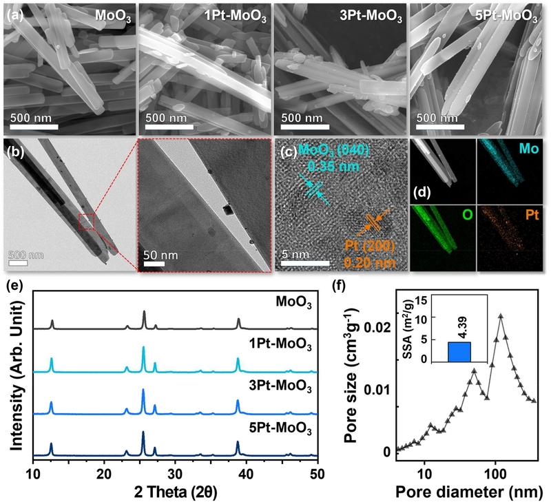
Structural and morphological characterization of Pt-MoO3 NRs. (a) FE-SEM images of MoO3, 1Pt-MoO3, 3Pt-MoO3, and 5Pt-MoO3. (b) TEM, (c) HR-TEM, and (d) EDS elemental mapping images of 3Pt-MoO3 NRs. (e) XRD patterns of all samples. (f) Pore size distribution and SSA (inset) of 3Pt-MoO3 NRs.
XRD patterns showed that all samples exhibited the orthorhombic MoO3 phase (JCPDS #05-0508) (Fig. 1(e)), consistent with the HR-TEM results. No noticeable shifts in the major diffraction peaks were observed with increasing Pt loading, suggesting that Pt was not incorporated into the MoO3 lattice. No diffraction peaks corresponding to Pt were observed, likely due to the low Pt content. These results a indicate that Pt decoration did not alter the crystal structure of the MoO3 NRs.
Pore-size analysis of 3Pt-MoO3 revealed pores spanning a broad size range from several to tens of nanometers (Fig. 1(f)), which can be attributed to void spaces formed between the assembled NRs. The SSA of 3Pt-MoO3 was 4.39 m2 g-1 (inset of Fig. 1(f)). These inter-rod void spaces provide pathways for gas transport through the sensing material, which is favorable for gas sensing.
3.2 Colorimetric Hydrogen Sensing Properties of Pt-MoO3 Sensors
The colorimetric H2-sensing properties of pure MoO3 and Pt-MoO3 sensors were evaluated using 5 vol% H2 at room temperature. Pure MoO3 initially appeared gray and showed little color change even after H2 exposure for more than 10 min (Fig. 2(a)). In contrast, all Pt-MoO3 sensors exhibited a clear color change to dark navy upon H2 exposure and did not return to their original color after H2 removal, demonstrating that Pt decoration is essential for the colorimetric H2 response. The initial color of the Pt-MoO3 sensors became progressively darker with increasing Pt loading.
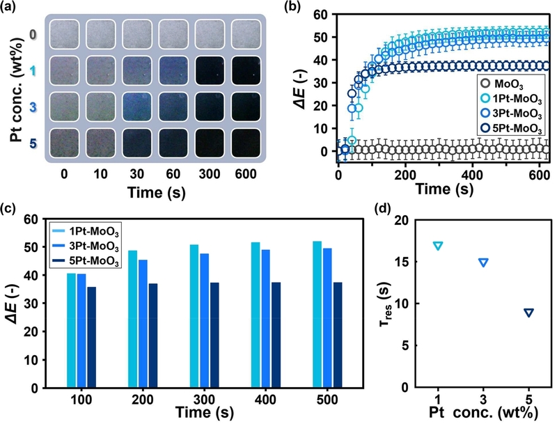
Colorimetric responses of Pt-MoO3 NRs to 5 vol% H2 at room temperature. (a) Time-dependent optical images. (b) Time-resolved changes in ΔE. (c) ΔE values at selected exposure times. (d) Response time (τres) as a function of Pt loading.
The color changes were quantitatively evaluated using the time-dependent ΔE (Fig. 2(b)). The results revealed contrasting effects of Pt loading on the magnitude and rate of the color change. The ΔE decreased in the order of 1Pt-MoO3, 3Pt-MoO3, and 5Pt-MoO3 over the measured exposure times (Fig. 2(c)), indicating that higher Pt loading resulted in a smaller color difference.
In contrast, the response time decreased with increasing Pt loading (Fig. 2(d)). Based on the time required to reach ΔE = 3, the response time decreased from approximately 17 s for 1Pt-MoO3 to 15 and 9 s for 3Pt-MoO3 and 5Pt-MoO3, respectively. Thus, increasing Pt loading resulted in a smaller color difference but a faster response, demonstrating its contrasting effects on the colorimetric sensing characteristics of Pt-MoO3.
3.3 Colorimetric Sensing Mechanism of Pt-MoO3 Sensors
To examine the H2-induced coloration mechanism of Pt-MoO3, the surface chemical states and optical properties of 3Pt-MoO3 were analyzed before and after H2 exposure. The Mo 3d XPS spectrum [17,18] of the pristine sample consisted of Mo6+ and Mo5+ components with relative fractions of 66.6 and 33.4%, respectively (Fig. 3(a)). The O 1s spectrum [19] showed lattice oxygen (OL, 81.8%), oxygen vacancies (OV, 16.4%), and surface hydroxyl species (OH, 1.8%) (Fig. 3(b)). Upon H2 exposure, the Mo5+ fraction increased to 71.4%, while the Mo6+ fraction decreased to 28.6% (Fig. 3(c)). Concurrently, the OV and OH fractions increased to 30.7 and 4.3%, respectively, while the OL fraction decreased to 65.0% (Fig. 3(d)). These changes indicate that H2 exposure reduces MoO3, accompanied by increases in oxygen vacancies and surface hydroxyl species.
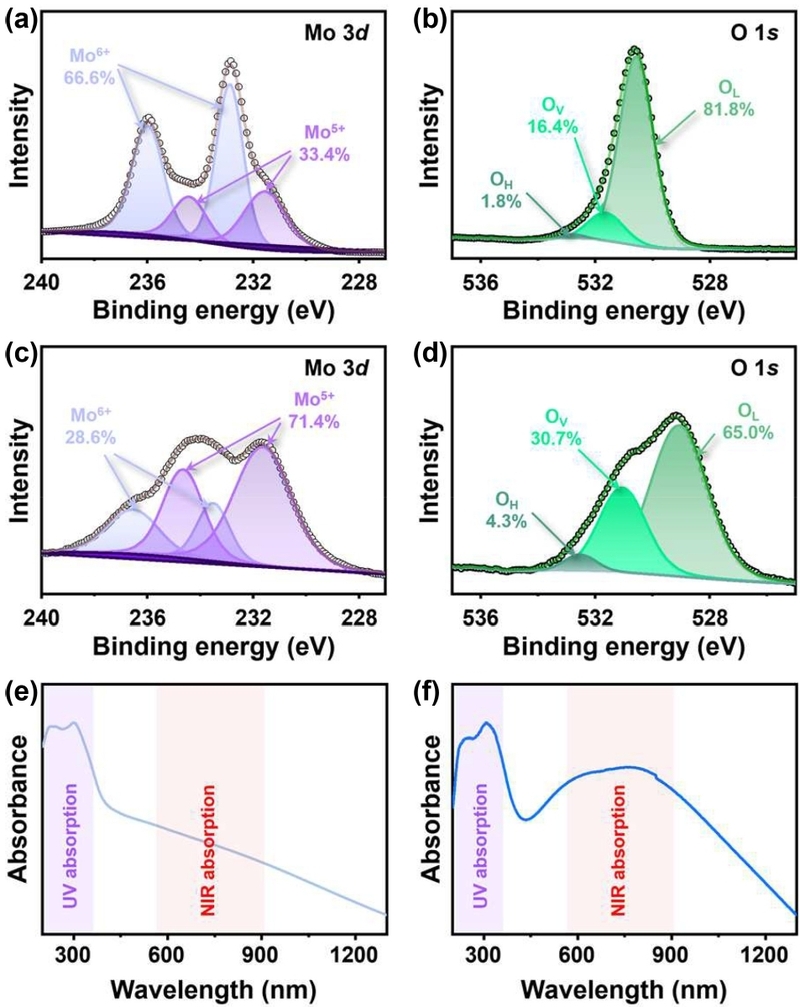
Surface chemical and optical analyses of 3Pt-MoO3 NRs. (a, b) Mo 3d and O 1s XPS spectra before H₂ exposure. (c, d) Mo 3d and O 1s XPS spectra after H₂ exposure. UV-Vis-NIR absorbance spectra (e) before and (f) after exposure to 5 vol% H₂.
The H2-induced chemical changes were accompanied by a pronounced change in optical absorption. Compared with the pristine sample (Fig. 3(e)), the H2-exposed sample showed substantially increased absorption across the visible and NIR regions (Fig. 3(f)). This increase can be attributed to reduced Mo species and to defect-related electronic states formed upon H2 exposure. In particular, intervalence charge transfer between adjacent Mo5+ and Mo6+ species can contribute to absorption in the visible region [20], accounting for the characteristic navy coloration. Taken together, the XPS and optical results indicate that H2 exposure reduces MoO3, increasing Mo5+ and oxygen vacancies and consequently altering its electronic structure and optical absorption. These changes produce the colorimetric H2 response of Pt-MoO3.
Pt nanoparticles facilitate this process by promoting H2 activation at room temperature [21]. Because direct H2 activation on MoO3 is limited, Pt promotes H2 dissociation and the transfer of activated hydrogen species to the MoO3 surface through hydrogen spillover [8]. This accounts for the negligible color change of pure MoO3 and the pronounced coloration of Pt-MoO3 upon H2 exposure. The faster response with increasing Pt loading can therefore be reasonably attributed to enhanced H2 activation and subsequent hydrogen transfer to MoO3.
In contrast, increasing Pt loading decreased ΔE. As shown in Fig. 2(a), the initial color became progressively grayer with increasing Pt loading. This change is likely associated with the optical contribution of Pt nanoparticles [22], as it differs from the characteristic navy coloration induced by H2 exposure. The darker initial color reduces the optical contrast between the initial and H2-exposed states, resulting in a smaller ΔE at higher Pt loadings. Therefore, increasing Pt loading accelerates the colorimetric response but decreases the resulting color difference. Considering these contrasting effects, 3Pt-MoO3 was selected for further evaluation because it retained a relatively large ΔE while exhibiting a shorter response time than 1Pt-MoO3 (Table 1).

Colorimetric H2-sensing characteristics of MoO3 NRs with different Pt loadings.
3.4 Gas Sensing Performance of the 3Pt-MoO3 Sensor
The H2-sensing performance of the selected 3Pt-MoO3 sensor was further evaluated under various conditions. Fig. 4(a) shows the time-dependent ΔE as the H2 concentration was decreased from 5 to 0.5 vol%. As the H2 concentration decreased, the color change became smaller and slower. Nevertheless, at 0.5 vol% H2, ΔE gradually exceeded the visible color-difference criterion (ΔE = 3) upon prolonged exposure. This demonstrates that 0.5 vol% H2 can be visually detected after prolonged exposure.
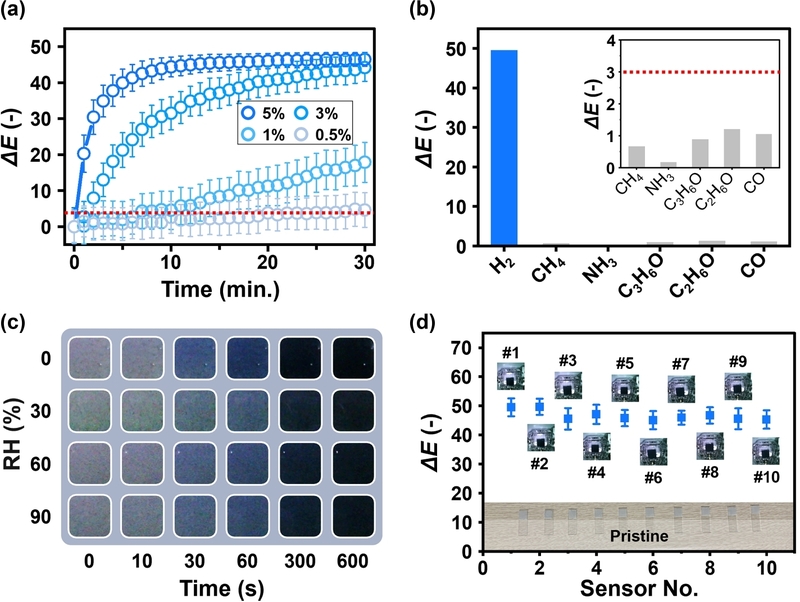
Colorimetric H2 sensing performance of 3Pt-MoO3 NRs. (a) Time-resolved ΔE changes at 0.5–5 vol% H2. (b) Selectivity against interfering gases. (c) Time-dependent optical images under 5 vol% H2 at RH 0–90%. (d) Sensor-to-sensor reproducibility under 5 vol% H2.
The selectivity of the 3Pt-MoO3 sensor was evaluated against CH₄, NH₃, acetone, ethanol, and CO (Fig. 4(b)). H2 produced a pronounced color change, whereas all interfering gases showed only minor changes, even after prolonged exposure. The ΔE values of the interfering gases remained below 3, demonstrating that the visible color change was highly selective to H2 under the tested conditions.
The effect of relative humidity (RH) on the H2 response was examined over an RH range of 0–90% (Fig. 4(c)). The sensor exhibited comparable color changes upon H2 exposure across the tested RH range, showing that its colorimetric response was not significantly affected by humidity. This humidity tolerance is favorable for H2 detection under varying environmental conditions.
Finally, sensor-to-sensor reproducibility was evaluated using ten 3Pt-MoO3 sensor pieces (Fig. 4(d)). All ten sensors exhibited similar ΔE values and visually consistent color changes upon H2 exposure. The small variation among the sensors confirms the reproducibility of the fabricated colorimetric sensors. Collectively, these results demonstrate that 3Pt-MoO3 maintains reliable colorimetric H2-sensing performance across different H2 concentrations, interfering gases, humidity conditions, and individual sensor units.
3.5 H2 Leakage Detection Using the 3Pt-MoO3 Colorimetric Sensor
The practical applicability of the 3Pt-MoO3 sensor was examined using a simple H2 leakage test (Fig. 5). The sensor film was attached near a gas-line fitting, which was slightly loosened to create a controlled leakage point, and 5 vol% H2 was subsequently supplied through the gas line. A commercial H2 sensor was used to monitor the surrounding
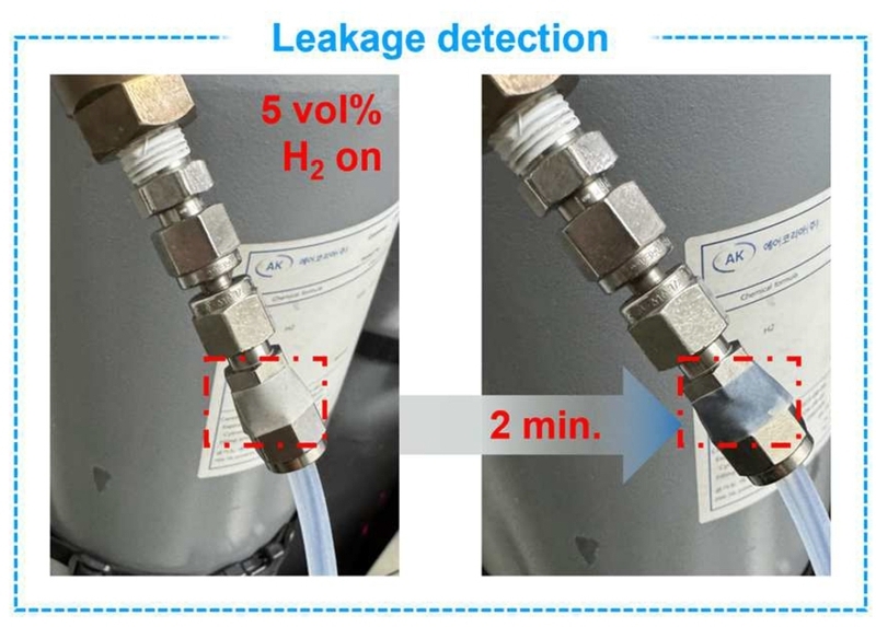
Demonstration of visual H2 leakage detection using a 3Pt-MoO3 sensor before and 2 min after 5 vol% H2 leakage from a gas-line fitting.
H2 concentration and ensure safety during the experiment. Before H2 exposure, the sensor appeared gray. Upon leakage from the fitting, the sensor region near the leakage point changed to dark navy within 2 min, allowing the leakage to be readily identified by visual inspection without additional signal readout. This demonstration shows that the 3Pt-MoO3 sensor can be directly attached near potential leakage points in H2 gas lines to provide a simple visual indication of leakage.
4. CONCLUSIONS
Pt-decorated MoO3 NRs with different Pt loadings were investigated for colorimetric H2 sensing at room temperature. Pt decoration was essential for inducing a clear color change upon H2 exposure. Increasing the Pt loading accelerated the response but decreased ΔE, showing contrasting effects on response kinetics and color difference. XPS and UV-Vis-NIR analyses indicated that the colorimetric response originates from H2-induced reduction of MoO3, accompanied by increased Mo5+ and oxygen vacancies. The faster response at higher Pt loadings was attributed to enhanced H2 dissociation and hydrogen spillover by Pt, whereas the decrease in color difference was associated with the darker initial color resulting from the optical contribution of Pt nanoparticles. Considering both color difference and response time, 3Pt-MoO3 was selected for further evaluation. It exhibited H2 selectivity, humidity tolerance, and sensor-to-sensor reproducibility and also enabled visual detection of H2 leakage in a gas-line leakage test. Overall, these results demonstrate the importance of Pt loading in determining the colorimetric sensing characteristics of Pt-MoO3 and provide useful guidance for developing colorimetric sensors for visual H2 leakage detection.
Acknowledgments
This work was supported by the Regional Innovation System & Education (RISE) program through the Jeonbuk RISE Center.
REFERENCES
-
K.T. Møller, T.R. Jensen, E. Akiba, H.-W. Li, Hydrogen – A sustainable energy carrier, Prog. Nat. Sci. Mater. Int. 27 (2017) 34–40.
[https://doi.org/10.1016/j.pnsc.2016.12.014]

-
T.M. Swager, T.N. Pioch, H. Feng, H.M. Bergman, S.-X.L. Luo, J.J. Valenza II, Critical Sensing Modalities for Hydrogen: Technical Needs and Status of the Field to Support a Changing Energy Landscape, ACS Sens. 9 (2024) 2205–2227.
[https://doi.org/10.1021/acssensors.4c00251]
-
G. Korotcenkov, S.D. Han, J.R. Stetter, Review of Electrochemical Hydrogen Sensors, Chem. Rev. 109 (2009) 1402–1433.
[https://doi.org/10.1021/cr800339k]
-
P. Ngene, T. Radeva, M. Slaman, R.J. Westerwaal, H. Schreuders, B. Dam, Seeing Hydrogen in Colors: Low-Cost and Highly Sensitive Eye Readable Hydrogen Detectors, Adv. Funct. Mater. 24 (2014) 2374–2382.
[https://doi.org/10.1002/adfm.201303065]
-
S. Mobtakeri, S. Habashyani, E. Gür, Highly responsive Pd-decorated MoO3 nanowall H₂ gas sensors obtained from in-situ-controlled thermal oxidation of sputtered MoS₂ films, ACS Appl. Mater. Interfaces 14 (2022) 25741–25752.
[https://doi.org/10.1021/acsami.2c04804]
-
S.S. Kalanur, I.-H. Yoo, H. Seo, Pd on MoO3 nanoplates as small-polaron-resonant eye-readable gasochromic and electrical hydrogen sensor, Sens. Actuators B Chem. 247 (2017) 357–365.
[https://doi.org/10.1016/j.snb.2017.03.033]
-
X. Sun, L. Hao, L. Chen, X. Guo, C. Han, J. Chen, et al., Spray deposition of colorimetric H₂ detector with Pd/MoO3 nanocomposites for rapid hydrogen leakage monitoring at room temperature, Appl. Surf. Sci. 599 (2022) 153878.
[https://doi.org/10.1016/j.apsusc.2022.153878]
-
J. Bai, J. Shang , J. Mei, X. Wang , C. Zhang , H. Kandambige, et al., Hydrogen Spillover Intensified by Pt Sites on Single-Crystalline MoO3 Interconnected Branches for Hydrogen Evolution, ACS Energy Lett. 8 (2023) 3868–3875.
[https://doi.org/10.1021/acsenergylett.3c01459]
-
Q. Liu, W. Xu, H. Huang, H. Shou, J. Low, Y. Dai, et al., Spectroscopic visualization of reversible hydrogen spillover between palladium and metal–organic frameworks toward catalytic semihydrogenation, Nat. Commun. 15 (2024) 2562.
[https://doi.org/10.1038/s41467-024-46923-3]
-
S.-I. Han, S.Y. Lee, L.T. Duy, H. Seo, Reversible gasochromic hydrogen sensing of mixed-phase MoO3 with multi-layered Pt/Ni/Pt catalyst, Int. J. Hydrogen Energy 46 (2021) 33339–33348.
[https://doi.org/10.1016/j.ijhydene.2021.07.091]
-
S. Singh, P. Bisht, J.P. Singh, Near-ambient NO2 sensing with vertical α-MoO3 nanorods synthesized by pulsed laser deposition, ACS Appl. Nano Mater. 8 (2025) 18624–18636.
[https://doi.org/10.1021/acsanm.5c03685]
-
H.-Y. Li, L. Huang, X.-X. Wang, C.-S. Lee, J.-W. Yoon, J. Zhou, et al., Molybdenum trioxide nanopaper as a dual gas sensor for detecting trimethylamine and hydrogen sulfide, RSC Adv. 7 (2017) 3680–3685.
[https://doi.org/10.1039/C6RA26280E]
-
D.T. Le, D.-S. Kim, G.-J. Oh, G.-S. Shin, T.V. Tran, B.-G. Ahn, et al., Dual single atoms (Pt, Ni) and PtNi alloy nanoparticles encapsulated N-doped carbon framework for durable ORR and HER electrocatalysts, Sustain. Mater. Technol. 41 (2024) e01068.
[https://doi.org/10.1016/j.susmat.2024.e01068]
-
D. Wu, Y. Shi, X. Liu, Q. Wang, C. Zhang, M. Hua, et al., Self-Cleaning Pd–TiO2–WO3 Heterostructure for High-Performance Hydrogen Gas Sensing, ACS Sens. 11 (2026) 6080–6092.
[https://doi.org/10.1021/acssensors.6c01069]
-
Z. Luo, R. Miao, T.D. Huan, I.M. Mosa, A.S. Poyraz, W. Zhong, et al., Mesoporous MoO3−x Material as an Efficient Electrocatalyst for Hydrogen Evolution Reactions, Adv. Energy Mater. 6 (2016) 1600528.
[https://doi.org/10.1002/aenm.201600528]
-
P. Xu, L. Dong, M. Neek-Amal, M.L. Ackerman, J. Yu, S.D. Barber, et al., Self-Organized Platinum Nanoparticles on Freestanding Graphene, ACS Nano 8 (2014) 2697–2703.
[https://doi.org/10.1021/nn406394f]
-
Z. Gu, M. Li, C. Chen, X. Zhang, C. Luo, Y. Yin, et al., Water-assisted hydrogen spillover in Pt nanoparticle-based metal–organic framework composites, Nat. Commun. 14 (2023) 5836.
[https://doi.org/10.1038/s41467-023-40697-w]
-
B. Li, W.L. Yim, Q. Zhang, L. Chen, A Comparative Study of Hydrogen Spillover on Pd and Pt Decorated MoO3(010) Surfaces from First Principles, J. Phys. Chem. C 114 (2010) 3052–3058.
[https://doi.org/10.1021/jp9098396]
-
Z. Lu, S.E. Saji, J. Langley, Y. Lin, Z. Xie, K. Yang, et al., Selective N2/H2O Adsorption onto 2D Amphiphilic Amorphous Photocatalysts for Ambient Gas-Phase Nitrogen Fixation, Appl. Catal. B Environ. 294 (2021) 120240.
[https://doi.org/10.1016/j.apcatb.2021.120240]
-
A. Borgschulte, O. Sambalova, R. Delmelle, S. Jenatsch, R. Hany, F. Nüesch, Hydrogen reduction of molybdenum oxide at room temperature, Sci. Rep. 7 (2017) 40761.
[https://doi.org/10.1038/srep40761]
-
K. Nishizawa, Y. Yamada, K. Yoshimura, Low-temperature chemical fabrication of Pt-WO3 gasochromic switchable films using UV irradiation, Sol. Energy Mater. Sol. Cells 170 (2017) 21–26.
[https://doi.org/10.1016/j.solmat.2017.05.058]
-
J. Reichstein, P. Groppe, N. Stockinger, C. Cuadrado Collados, M. Thommes, S. Wintzheimer, et al., Safety through visibility: Tracing hydrogen in colors with highly customizable and flexibly applicable supraparticle additives, Adv. Mater. Technol. 9 (2024) 2400441.
[https://doi.org/10.1002/admt.202400441]