
Piezoelectric Biomolecules for Sustainable and Biointegrated Sensors

ⓒ The Korean Sensors Society
This is an Open Access article distributed under the terms of the Creative Commons Attribution Non-Commercial License (https://creativecommons.org/licenses/by-nc/3.0/) which permits unrestricted non-commercial use, distribution, and reproduction in any medium, provided the original work is properly cited.
Abstract
Piezoelectric biomolecules provide a materials basis for soft, degradable, and self-powered sensing systems by combining electromechanical transduction with biological structure and chemistry. Their piezoelectricity arises from ordered molecular and supramolecular polarities, including zwitterionic amino acid crystals, hydrogen-bonded peptide assemblies, polar protein and polysaccharide hierarchies, and engineered biological templates. This review examines how these structural motifs are translated into sensor materials and biointegrated devices. We first classify representative piezoelectric biomolecules by their polarization origins, then discuss assembly and fabrication strategies that improve macroscopic output through phase selection, nanoconfinement, fibril or fiber alignment, and electric-field-guided growth. Recent sensor demonstrations are summarized in wearable pressure sensing, acoustic and vibration detection, humidity monitoring, biodegradable physiological sensing, and implantable soft-tissue monitoring. Across these examples, the key design problem is not simply identifying polar biomolecules, but preserving, amplifying, and coupling their polarity across molecular, mesoscale, and device length scales under realistic operating conditions.
Keywords:
Piezoelectric biomolecules, Self-Assembly, Biodegradable sensors, Biointegrated electronics1. INTRODUCTION
Piezoelectric materials convert mechanical deformation into electrical polarization and form the foundation of sensors, actuators, acoustic transducers, and energy-harvesting devices. Conventional ceramic, oxide, and fluorinated-polymer piezoelectrics offer mature processing and strong electromechanical coupling, but their stiffness, processing requirements, limited degradability, and long-term persistence can be mismatched with soft or transient bioelectronics [1-3].
These limitations have encouraged the search for soft and bio-derived piezoelectric materials. Piezoelectric biomolecules are built from amino acids, peptides, proteins, polysaccharides, and biological templates, and can combine electromechanical transduction with aqueous processing, biocompatibility, biodegradability, and chemical functionality [4-6]. Such features are useful not only for passive sensing but also for biointerfaces in which polarization can influence cellular activity or tissue responses [7].
The material scope now spans several structural levels. Amino acid crystals such as glycine and alanine reveal how zwitterionic dipoles become piezoelectric when crystal symmetry and domain orientation are controlled [8,9]. Peptide assemblies, including Diphenylalanine (FF) nanotubes and sequence-programmed peptide crystals, show how hydrogen bonding, aromatic packing, and side-chain chemistry tune supramolecular polarization [10,11]. Proteins, polysaccharides, and M13 virus extend the same concept to collagen helices, silk β-sheets, nanocellulose crystals, and genetically tunable viral templates [12-17].
A recurring bottleneck is the need to preserve local polarity over device-relevant length scales. Random crystal orientation, mixed polymorphs, hydration-induced screening, weak stress transfer, and poor electrode coupling can suppress output even when the molecular building block is polar. Recent processing routes therefore focus on stabilizing active phases and aligning dipoles through meniscus-guided crystallization, nanoconfinement, magnetic-field alignment, electrospinning, and electric-field-assisted growth [8,18-22].
These advances have enabled biomolecular piezoelectric sensors for pressure and force sensing, acoustic and vibration readout, humidity monitoring, biodegradable physiological sensing, and implantable soft-tissue monitoring [23-27]. This review follows the structure-processing-application pathway: first, classifying piezoelectric biomolecules by polarization origin, then discussing alignment and self-assembly strategies, and finally comparing representative sensing devices.
2. STRUCTURAL ORIGINS AND CLASSIFICATION OF PIEZOELECTRIC BIOMOLECULES
Piezoelectric biomolecules can be classified by the structural level at which polarity is organized, from small molecular crystals to supramolecular assemblies, macromolecular hierarchies, and biological templates. In all cases, electromechanical coupling requires local dipoles to be arranged in a non-centrosymmetric or anisotropic manner so that deformation changes the net polarization rather than cancelling it. The relevant dipoles arise from zwitterionic groups, polar peptide bonds, hydrogen-bonded backbones, sugar-chain polarity, charged residues, or ordered protein surfaces. Fig. 1 uses representative structures to illustrate these families, but the central point is the underlying structural origin of piezoelectricity: crystal symmetry in amino acids, polar self-assembly in peptides, secondary structure and fibrillar anisotropy in proteins, oriented hydrogen-bonded crystalline domains in polysaccharides, and surface-charge alignment in biological templates [1-6]. These structural motifs also determine how the active layer is later coupled to devices, as seen in ultrathin peptide monolayers, glycine-containing force sensors, and silk-based implantable constructs [23-25]. Quantitative coefficients and conventional-material benchmarks are consolidated in Table 1 at the end of Section 2. The corresponding processing routes, performance ranges, advantages, and limitations are compared in Table 2 at the end of Section 3.
2.1 Amino Acids
Amino acids represent the smallest class of piezoelectric biomolecules (Fig. 1(a)). Their intrinsic polarity is mainly carried by zwitterionic NH3+/COO- groups, but molecular dipoles alone are insufficient; the molecules must pack into a non-centrosymmetric crystal so that their dipoles are not cancelled by inversion symmetry. This explains why different polymorphs of the same amino acid can behave very differently. In glycine, for example, the centrosymmetric α phase is essentially non-piezoelectric, whereas β- and γ-glycine are polar and can exhibit measurable shear or longitudinal responses [8,9]. Reported values also show the importance of packing and orientation: β-glycine has been associated with a large shear response, while γ-glycine is more stable and can provide out-of-plane responses on the order of several pC N-1 when processed into films or composites [8-10]. Alanine further shows that chirality and racemic packing affect the final crystal symmetry and dipole arrangement rather than acting as simple one-parameter predictors [9,28]. Mechanically engineered amino-acid crystals, supramolecularly engineered assemblies, and polymer-supported alanine polycrystals extend this principle by improving crystallinity, morphology, orientation, and stress transfer while retaining device compatibility [26,29,30]. Thus, amino-acid piezoelectricity is best described as zwitterionic dipoles organized by crystal symmetry, with polymorphism, hydrogen bonding, chirality, and domain orientation acting as coupled control factors.
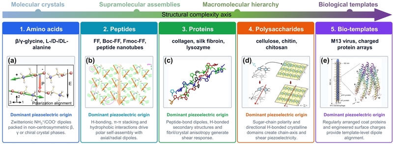
Structural Origins and Classification of Piezoelectric Biomolecules. (a) β-glycine. Adapted from Ref. [8]. (b) phenylalanine-centered hydrophobic tripeptide crystals. Adapted from Ref. [10]. (c) collagen triple helix. Adapted from Ref. [14]. (d) Iβ-nanocellulose. Adapted from Ref. [16]. (e) M13 bacteriophage. Adapted from Ref. [17].
2.2 Peptides
Peptides add supramolecular programmability to biomolecular piezoelectricity (Fig. 1(b)). Their piezoresponse is not governed only by the polarity of an isolated peptide bond, but by how many peptide molecules assemble into ordered architectures through hydrogen bonding, aromatic π-π stacking, hydrophobic interactions, electrostatic interactions, and side-chain packing. Diphenylalanine (FF) is the benchmark example: FF can form nanotubes, rods, and crystalline assemblies in which axial or radial polarization emerges from the packing geometry [12,31]. Piezoresponse force microscopy and device studies have shown strong shear responses in FF nanotubes and voltage/current outputs from aligned peptide nanogenerators, highlighting the need for both molecular polarity and macroscopic orientation [32]. More broadly, sequence-programmed peptide crystals and collagen-mimicking peptide assemblies demonstrate that side-chain chemistry, aromaticity, heterochirality, hydrogen-bond geometry, and crystallization pathway can tune symmetry and polarization [11,13]. Peptide piezoelectricity therefore originates from polar self-assembly into non-centrosymmetric supramolecular structures, with sequence design and alignment determining whether the local dipoles add constructively.
2.3 Proteins
Proteins provide a higher level of structural hierarchy (Fig. 1(c)). Their active dipoles are distributed across peptide bonds, polar side chains, and hydrogen-bonded secondary structures, but a macroscopic response requires these local polar units to be embedded in anisotropic fibrils, fibers, crystals, or tissue matrices. Collagen is the classical biological example. Its triple helix contains aligned amide groups and an extended hydrogen-bonded backbone, and the organization of triple helices into fibrils and fibers produces direction-dependent shear piezoelectricity in collagen-rich tissues such as bone and tendon [14,33,34]. Reported collagen and bone responses are generally modest compared with optimized small-molecule crystals, but they are biologically important because the electromechanical response is integrated with load-bearing extracellular matrices [33,34]. Silk fibroin follows a related but distinct mechanism, where oriented β-sheet-rich domains and chain alignment generate polar order that can be strengthened by drawing or regeneration [15]. Globular protein crystals, such as lysozyme, further demonstrate that proteins need not be fibrous to be piezoelectric if their crystal packing is polar [35]. Overall, protein piezoelectricity arises from peptide-bond polarity amplified by secondary-structure order, crystal packing, and hierarchical anisotropy.
2.4 Polysaccharides
Polysaccharides generate piezoelectricity through polar sugar chains and directional hydrogen-bonded crystalline domains (Fig. 1(d)). In cellulose, β-1,4-linked glucan chains form crystalline nanodomains in which hydroxyl groups, glycosidic bonds, and chain-axis polarity are arranged anisotropically. The resulting response is often shear-dominated and strongly depends on nanocrystal or nanofibril orientation, film morphology, hydration, and the deformation direction [16,36,37]. Cellulose nanocrystal and nanofibril films have therefore become useful model systems for studying how renewable fibrillar networks convert mechanical strain into electrical signals [36,37]. Chitin and chitosan follow the same structural logic but introduce acetamide or amine groups that modify hydrogen bonding, polarity, crystallinity, and water interaction [38]. β-Chitin nanofibrils isolated from deep-sea tubeworms further demonstrate that native crystallinity and c-axis alignment can support low-frequency mechano-electric coupling without external poling [39]. In this class, the key origin is not a single permanent dipole, but the collective deformation of oriented polar chains and hydrogen-bonded crystalline domains. This also explains why processing, alignment, crystallinity, and humidity control are central to polysaccharide-based piezoelectric devices.
2.5 Biological Templates
Biological templates represent a distinct route in which long-range order is supplied by an organized biological object rather than by a small-molecule or polymer crystal (Fig. 1(e)). M13 bacteriophage is the most widely studied example. Its rod-like body contains thousands of coat proteins arranged around the viral genome, creating an ordered protein shell with directional surface charges and dipoles. Genetic engineering can introduce charged residues and tune the surface charge distribution, while alignment of viral films determines whether these nanoscale dipoles produce a macroscopic response [17,40]. Virus-based nanogenerators have shown that engineered and aligned M13 films can generate electrical output under mechanical deformation, and later work has clarified how charge distribution, assembly, and interfacial effects contribute to the measured response [40,41]. The piezoelectric origin of such bio-templates is therefore regular surface-charge distribution combined with template-level orientation, with genetic modification and film alignment serving as the main design variables.
To consolidate these structural classes, Table 1 compares representative tensor components, sample forms, and measurement methods with PVDF and PZT benchmarks.

Representative piezoelectric coefficients of biomolecular systems and conventional benchmarks
3. FABRICATION AND SELF-ASSEMBLY ENGINEERING OF PIEZOELECTRIC BIOMOLECULES
3.1 Meniscus-Driven Self-Assembly
Meniscus-driven self-assembly uses a moving liquid contact line to couple evaporation, solute transport, nucleation, and crystal orientation. In β-glycine coating (Fig. 2(a)), pulling speed and concentration gradients determine whether rapid local supersaturation kinetically captures the metastable β phase or whether the system relaxes toward the thermodynamically stable α phase [18]. The free-energy and β-to-α conversion schemes in Fig. 2(a) therefore illustrate a general processing rule for biomolecular crystals: the polar phase must be selected before it transforms or grows into randomly oriented domains. Similar meniscus- and interface-guided approaches have been used to organize bio-organic thin films and γ-glycine-based flexible films, where substrate chemistry, evaporation rate, and interfacial confinement regulate polymorph selection, grain orientation, and stress transfer [19,49]. For amino-acid piezoelectrics, this coating route is especially useful because it links large-area film formation with the kinetic stabilization of a polar molecular crystal.
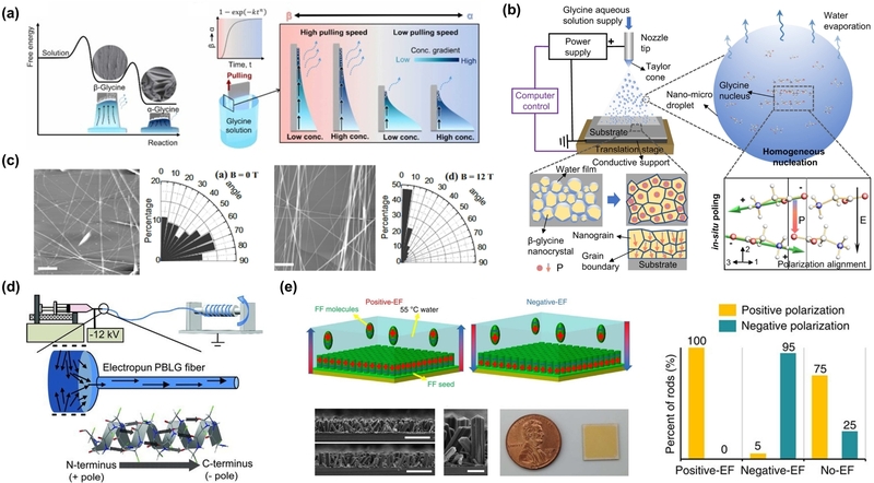
Fabrication and self-assembly engineering of piezoelectric biomolecules. (a) Meniscus-driven self-assembly of β-glycine. Adapted from Ref. [18]. (b) Electrospray-assisted nanoconfinement synthesis of β-glycine nanocrystalline films. Adapted from Ref. [8]. (c) Magnetic-field alignment of FF nanotube. Adapted from Ref. [20]. (d) Electrospinning aligns α-helical poly (amino acid) chains. Adapted from Ref. [21]. (e) Electric-field-assisted FF peptide self-assembly. Adapted from Ref. [12].
3.2 Electrospray-Assisted Nanoconfinement Synthesis
Electrospray-assisted nanoconfinement combines droplet generation, fast solvent evaporation, confined nucleation, and electrical bias. In β-glycine films (Fig. 2(b)), charged nano- to microdroplets land on the substrate, where a thin water layer promotes homogeneous nucleation while in situ poling biases the orientation of growing nanocrystals [8]. Fig. 2(b) shows this sequence from droplet formation to nanograin assembly and polarization alignment. Beyond this example, nanoconfinement is broadly useful for biomolecular piezoelectrics because it suppresses uncontrolled crystal growth, increases phase uniformity, and enables polar domains to form directly in thin-film geometries. This concept is also consistent with wafer-scale bio-organic thin films and interface-induced glycine films, in which confined crystallization and substrate interactions stabilize polar phases and improve device-level output [19,49].
3.3 Magnetic-Field Alignment
Magnetic-field alignment addresses orientational disorder in anisotropic supramolecular assemblies. FF nanotubes contain aromatic groups and ordered hydrogen-bonded backbones, making their long axes sensitive to strong magnetic fields. In Fig. 2(c), without an applied field, the angular distribution is broad; under 12 T, the nanotube population becomes concentrated near a preferred direction, and the SEM image shows a more aligned network [20]. This result extends beyond FF because many peptide and protein assemblies are mechanically and electrically anisotropic. Their output depends on whether the tube, fiber, or fibril axis is aligned with stress direction and electrode configuration. Magnetic alignment is therefore a useful model strategy for proving the role of supramolecular orientation, even if scalable device manufacturing may rely more on coating, shearing, or electric-field methods.
3.4 Electrospinning
Electrospinning provides simultaneous fiber formation and molecular alignment. The high electric field and elongational flow stretch chains, guide solvent removal, and produce continuous fibers that efficiently transfer stress. Electrospun PBLG fibers in Fig. 2(d) show a preferred molecular orientation from the N-terminus positive pole toward the C-terminus negative pole, explaining their permanent polarity without high-temperature poling [21]. The same fiber-processing logic has been applied to peptide nanotubes embedded in electrospun polymer fibers and to biodegradable amino acid or peptide composite fibers, where the polymer matrix improves flexibility while the polar biomolecular phase supplies electromechanical conversion [50,51]. A conventional-polymer comparison reinforces the same processing principle: mechanically stretching electrospun PVDF non-woven fabrics increased β-phase content and electrical output by improving fiber orientation [52]. Compared with flat crystals, electrospun mats are better suited for wearable and implantable devices because they are porous, conformal, and mechanically robust under repeated bending.
3.5 Electric-Field Alignment
Electric-field alignment is particularly effective when applied during nucleation and growth. In FF peptide assembly (Fig. 2(e)), a positive electric field produces nearly 100% positively polarized rods, a negative electric field produces about 95% negatively polarized rods, and no field leaves a mixed distribution of roughly 75% positive and 25% negative rods [12]. These data in Fig. 2(e) explain why the route improves macroscopic output: field-guided growth increases the number of domains that contribute to the same polarization direction. Related electric-field-assisted peptide self-assembly and solvent-mediated unidirectional nanotube growth show that molecular dipoles, seed crystals, solvent environment, and electrode geometry can all be used to program peptide polarity [22,53]. More generally, field alignment provides a direct way to convert the intrinsic polarity of peptide assemblies into a device-scale polar film before random domains are locked in.
Across these routes, the practical choice is governed by the same coupled variables—phase selection, dipole alignment, stress transfer, scale-up, and environmental stability—which are summarized in Table 2. The fabrication routes discussed above address different bottlenecks, but their performance depends on the structural class being processed. Table 2 therefore links each biomolecular category to its main processing routes, representative coefficient range, strengths, and practical limitations.
Concise comparison of biomolecular categories, fabrication routes, strengths, and limitations.
4. PIEZOELECTRIC BIOMOLECULAR SENSORS FOR BIOINTEGRATED AND SELF-POWERED SENSING APPLICATIONS
4.1 Wearable Pressure and Force Sensing
Wearable pressure and force sensors require thin active layers that respond to gentle touch, bending, and repeated contact. Self-assembled oligopeptide monolayers provide an ultrathin polar platform in which molecular dipoles are intrinsic to the assembly [23]. In the wearable peptide sensor (Fig. 3(a)), pressing or bending the flexible device produces clear current and voltage pulses, with stronger deformation generating larger voltage signals on the order of volts. The charge coefficient reaches about 9.8 pC N-1, while the voltage coefficient can reach 2 V m N-1, a useful feature for lightweight self-powered force readout [23]. Other biodegradable force sensors use glycine-chitosan composites or mechanically annealed amino acid crystals to combine crystal-domain control with soft matrices, expanding this application from molecular monolayers to flexible degradable pressure devices [26,54].
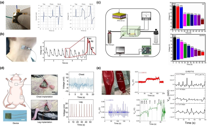
Piezoelectric biomolecular sensors for biointegrated and self-powered sensing applications. (a) Wearable pressure and force sensing. Adapted from Ref. [23]. (b) Acoustic and vibration sensing. Adapted from Ref. [24]. (c) Humidity and environmental sensing. Adapted from Ref. [27]. (d) Biodegradable physiological motion sensing. Adapted from Ref. [26]. (e) Implantable soft-tissue monitoring. Adapted from Ref. [25].
4.2 Acoustic and Vibration Sensing
Acoustic and vibration sensing relies on fast electromechanical response to small dynamic deformation. The β-glycine/alginate film (Fig. 3(b)) is attached to the neck and produces repeated millivolt-scale voltage peaks during vocal or throat motion [24]. The enlarged waveform contains multiple peaks within each event, indicating that the device can resolve complex mechanical vibrations rather than only detecting a single pressure pulse. This is relevant to voice, swallowing, and soft acoustic interfaces, where waveform shapes carry physiological information. Peptide nanotube substrate sensors provide a complementary route: sound waves activate the peptide piezoelectric layer and generate electrical signals, connecting supramolecular peptide polarity to acoustic readout [55].
4.3 Humidity and Environmental Sensing
Humidity and environmental sensors exploit the water sensitivity of biopolymers. In the chitosan/PVA system (Fig. 3(c)), a piezoelectric nanogenerator powers a humidity-sensitive CS-PVA film, and the output changes systematically as relative humidity increases from 21% to 89% [27]. The voltage response decreases from approximately 15 V at low humidity to about 2 V at 89% RH, reflecting water adsorption, swelling, and changes in charge transport in the hydrophilic sensing layer. The stepwise response allows battery-free humidity discrimination. Collagen and cellulose devices show related hydration-dependent behavior, where water can either be the analyte or an interference that screens polarization and alters mechanical stiffness [36,37,56]. More broadly, recent humidity-sensor literature identifies sensitivity, linearity, operating range, response/recovery time, and long-term stability as shared evaluation criteria across material platforms [57].
4.4 Biodegradable Physiological Motion Sensing
Biodegradable physiological motion sensors must record low-frequency body movements while minimizing long-term material retention. Mechanically annealed amino-acid crystals provide degradable force sensors with improved sensitivity through enhanced packing and domain orientation [26]. In the biodegradable physiological sensor (Fig. 3(d)), implanted devices record periodic signals from chest and leg motion: chest implantation gives repeated millivolt-scale peaks, while leg motion produces larger spikes approaching tens of millivolts. The difference reflects the larger deformation amplitude and loading direction associated with limb movement. Earlier biodegradable piezoelectric force sensors and nanofiber-based transducers enabled transient biomedical electromechanical monitoring, while the amino acid crystal approach connects physiological sensing directly to molecular crystal engineering [58,59].
4.5 Implantable Soft-Tissue Monitoring
Implantable soft-tissue monitoring requires intimate contact with curved organs, mechanical compliance, and stable signal output in wet physiological environments. The 3D-printed silk-based bioresorbable piezoelectric self-adhesive holey structures (Fig. 3(e)) address these requirements through a patterned silk composite that conforms to tissue and maintains electrical connection [25]. The device records dynamic voltage changes under pressure steps ranging from several to more than 20 mbar and detects rupture events, demonstrating its ability to track both gradual deformation and abrupt mechanical failure. In vitro traces after 7, 14, and 21 days at 37 °C and pH 7.4 retain periodic voltage signals, indicating operation over degradation-relevant time windows. Skin-inspired tactile arrays and ultrastable piezoelectric biomaterial fabrics further show that electrode routing, encapsulation, and mechanical robustness become decisive when soft biomolecular sensors are integrated with living tissues [60,61].
5. CHALLENGES AND OUTLOOK
Piezoelectric biomolecules now provide a credible materials basis for soft and degradable sensors, but reproducibility remains the main barrier. The β-glycine nanocrystalline-film study [8] provides a useful model because phase identification and orientation mapping were combined with piezoresponse force microscopy, ferroelectric switching, and quasi-static coefficient measurements. The meniscus-stabilized β-glycine study [18] likewise linked processing conditions to polymorph selection and anisotropic response, while the silk study [15] correlated β-sheet content and crystalline orientation with the shear coefficient. Together, these cases show that a convincing benchmark should connect the reported coefficient to phase fraction, crystal orientation, domain polarity, hydration state, modulus, dielectric properties, and processing history. Studies that report only a peak voltage or current cannot establish whether an improvement originates from the biomolecule, device geometry, stress concentration, or the measurement circuit. Temperature should be included in this qualification because conventional piezoelectric materials already show material-dependent trade-offs among thermal depolarization, sensitivity, stiffness, and cyclic resilience [62].
Device-level evaluation also needs clearer standards. As Table 1 makes explicit, coefficients obtained by piezoresponse force microscopy, Berlincourt-type direct measurements, and device-level charge sensitivity are useful but not strictly interchangeable. The wearable, acoustic, humidity, biodegradable, and implantable demonstrations in Refs. [23-27] use different force ranges, loading geometries, frequencies, active areas, and electrical loads, which makes ranking by open-circuit voltage alone unreliable. Future studies should report calibrated force or stress, active area and thickness, loading rate and frequency, electrical boundary conditions, response time, cycling stability, signal-to-noise ratio, and uncertainty. Hydrophilic systems further require controlled-humidity or wet-state testing because water adsorption can simultaneously alter polarization screening, dielectric loss, and stiffness, as illustrated by the chitosan/PVA, collagen, and cellulose examples [27,36,37,56].
The most promising direction is hybrid biointegrated design. Amino acid or peptide crystals can provide high polarization, while protein and polysaccharide matrices supply compliance, processability, and biofunctionality; γ-glycine-polymer films [49], glycine-chitosan devices [54], and silk-based constructs [25] illustrate this complementary strategy. Biological templates can additionally impose long-range order, and scalable alignment, stress-focusing microstructures, soft electrodes, and low-power electronics can translate that order into robust signals. For implantable or transient systems [25,58-61], however, electromechanical stability should be evaluated together with sterilization tolerance, encapsulation lifetime, cytotoxicity, degradation products, inflammatory response, and biofouling. This combined materials-device-biological framework is needed to move beyond biodegradable substitutes for inorganic piezoelectrics toward sensors purpose-designed for transient physiological monitoring, self-powered environmental readout, and mechanically active biointerfaces.
Acknowledgments
This work was supported by the National Research Foundation of Korea (NRF) funded by the Ministry of Science and ICT (RS-2025-00560648). The Ministry of Science and ICT (26-InnoCORE-03 and N10250154).
REFERENCES
-
M.T. Chorsi, E.J. Curry, H.T. Chorsi, R. Das, J. Baroody, P.K. Purohit, et al., Piezoelectric Biomaterials for Sensors and Actuators, Adv. Mater. 31 (2019) 1802084.
[https://doi.org/10.1002/adma.201802084]

-
S. Chen, X. Tong, Y. Huo, S. Liu, Y. Yin, M.-L. Tan, et al., Piezoelectric Biomaterials Inspired by Nature for Applications in Biomedicine and Nanotechnology, Adv. Mater. 36 (2024) 2406192.
[https://doi.org/10.1002/adma.202406192]
-
Z. Zhang, Z. Wang, X. Li, Y. Zheng, Z. Yang, Design and Manufacturing of Piezoelectric Biomaterials for Bioelectronics and Biomedical Applications, Chem. Rev. 125 (2025) 9875–9929.
[https://doi.org/10.1021/acs.chemrev.5c00399]
-
R. Wang, J. Sui, X. Wang, Natural Piezoelectric Biomaterials: A Biocompatible and Sustainable Building Block for Biomedical Devices, ACS Nano 16 (2022) 17708–17728.
[https://doi.org/10.1021/acsnano.2c08164]
-
H. Yuan, P. Han, K. Tao, S. Liu, E. Gazit, R. Yang, Piezoelectric Peptide and Metabolite Materials, Research 2019 (2019) 9025939.
[https://doi.org/10.34133/2019/9025939]
-
K. Kapat, Q.T.H. Shubhra, M. Zhou, S. Leeuwenburgh, Piezoelectric Nano-Biomaterials for Biomedicine and Tissue Regeneration, Adv. Funct. Mater. 30 (2020) 1909045.
[https://doi.org/10.1002/adfm.201909045]
-
H. Guo, D.-M. Lee, P. Zhao, S.-H. Kim, I. Hyun, B.-J. Park, et al., Cell activity manipulation through optimizing piezoelectricity and polarization of diphenylalanine peptide nanotube-based nanocomposite, Chem. Eng. J. 468 (2023) 143597.
[https://doi.org/10.1016/j.cej.2023.143597]
-
Z. Zhang, X. Li, Z. Peng, X. Yan, S. Liu, Y. Hong, et al., Active self-assembly of piezoelectric biomolecular films via synergistic nanoconfinement and in-situ poling, Nat. Commun. 14 (2023) 4094.
[https://doi.org/10.1038/s41467-023-39692-y]
-
S. Guerin, A. Stapleton, D. Chovan, R. Mouras, M. Gleeson, C. McKeown, et al., Control of piezoelectricity in amino acids by supramolecular packing, Nat. Mater. 17 (2018) 180–186.
[https://doi.org/10.1038/nmat5045]
-
M.T. Chorsi, T.T. Le, F. Lin, T. Vinikoor, R. Das, J.F. Stevens, et al., Highly piezoelectric, biodegradable, and flexible amino acid nanofibers for medical applications, Sci. Adv. 9 (2023) eadg6075.
[https://doi.org/10.1126/sciadv.adg6075]
-
X. Yang, S. Liu, H. Lu, Y. Wang, H. Zhang, W. Ji, et al., Peptide Sequence Programmed Piezoelectric Response by Supramolecular Self-Assembly, Advanced Science 13 (2026) e15237.
[https://doi.org/10.1002/advs.202515237]
-
V. Nguyen, R. Zhu, K. Jenkins, R. Yang, Self-assembly of diphenylalanine peptide with controlled polarization for power generation, Nat. Commun. 7 (2016) 13566.
[https://doi.org/10.1038/ncomms13566]
-
S. Bera, S. Guerin, H. Yuan, J. O'Donnell, N.P. Reynolds, O. Maraba, et al., Molecular engineering of piezoelectricity in collagen-mimicking peptide assemblies, Nat. Commun. 12 (2021) 2634.
[https://doi.org/10.1038/s41467-021-22895-6]
-
J. Hartmann, M. Zacharias, Mechanism of collagen folding propagation studied by Molecular Dynamics simulations, PLOS Comput. Biol. 17 (2021) e1009079.
[https://doi.org/10.1371/journal.pcbi.1009079]
-
T. Yucel, P. Cebe, D.L. Kaplan, Structural Origins of Silk Piezoelectricity, Adv. Funct. Mater. 21 (2011) 779–785.
[https://doi.org/10.1002/adfm.201002077]
-
Y. García, Y.B. Ruiz-Blanco, Y. Marrero-Ponce, C.M. Sotomayor-Torres, Orthotropic Piezoelectricity in 2D Nanocellulose, Sci. Rep. 6 (2016) 34616.
[https://doi.org/10.1038/srep34616]
-
H. Kim, S.-W. Lee, Molecular Mechanisms and Enhancement of Piezoelectricity in the M13 Virus, Adv. Funct. Mater. 34 (2024) 2407462.
[https://doi.org/10.1002/adfm.202407462]
-
Y. Ryu, C. Lee, P. Zhao, J. Yeon, J. Choi, T.O. Ibrahim, et al., Large-area kinetic stabilization of metastable β-glycine via meniscus-driven self-assembly with anisotropic piezoelectricity, Nano Energy 154 (2026) 112005.
[https://doi.org/10.1016/j.nanoen.2026.112005]
-
F. Yang, J. Li, Y. Long, Z. Zhang, L. Wang, J. Sui, et al., Wafer-scale heterostructured piezoelectric bio-organic thin films, Science 373 (2021) 337–342.
[https://doi.org/10.1126/science.abf2155]
-
R.J.A. Hill, V.L. Sedman, S. Allen, P. Williams, M. Paoli, L. Adler-Abramovich, et al., Alignment of Aromatic Peptide Tubes in Strong Magnetic Fields, Adv. Mater. 19 (2007) 4474–4479.
[https://doi.org/10.1002/adma.200700590]
-
D. Farrar, K. Ren, D. Cheng, S. Kim, W. Moon, W.L. Wilson, et al., Permanent Polarity and Piezoelectricity of Electrospun α-Helical Poly(α-Amino Acid) Fibers, Adv. Mater. 23 (2011) 3954–3958.
[https://doi.org/10.1002/adma.201101733]
-
Y. Su, J. Liu, D. Yang, W. Hu, X. Jiang, Z.L. Wang, et al., Electric Field-Assisted Self-Assembly of Diphenylalanine Peptides for High-Performance Energy Conversion, ACS Mater. Lett. 5 (2023) 2317–2323.
[https://doi.org/10.1021/acsmaterialslett.3c00664]
-
C.A. Petroff, G. Cassone, J. Šponer, G.R. Hutchison, Intrinsically Polar Piezoelectric Self-Assembled Oligopeptide Monolayers, Adv. Mater. 33 (2021) 2007486.
[https://doi.org/10.1002/adma.202007486]
-
Q. Lin, Y. Zhang, L. Chen, H. Zhang, C. An, C. Li, et al., Glycine/alginate-based piezoelectric film consisting of a single, monolithic β-glycine spherulite towards flexible and biodegradable force sensor, Regener. Biomater. 11 (2024) rbae047.
[https://doi.org/10.1093/rb/rbae047]
-
I. Chiesa, C. De Maria, M.R. Ceccarini, L. Mussolin, R. Coletta, A. Morabito, et al., 3D Printing Silk-Based Bioresorbable Piezoelectric Self-Adhesive Holey Structures for In Vivo Monitoring on Soft Tissues, ACS Appl. Mater. Interfaces 14 (2022) 19253–19264.
[https://doi.org/10.1021/acsami.2c04078]
-
Y. Cheng, J. Xu, L. Li, P. Cai, Y. Li, Q. Jiang, et al., Boosting the Piezoelectric Sensitivity of Amino Acid Crystals by Mechanical Annealing for the Engineering of Fully Degradable Force Sensors, Advanced Science 10 (2023) 2207269.
[https://doi.org/10.1002/advs.202207269]
-
Zeeshan, A.M. Soomro, S. Cho, Design and Fabrication of a Robust Chitosan/Polyvinyl Alcohol-Based Humidity Sensor energized by a Piezoelectric Generator, Energies 15 (2022) 7609.
[https://doi.org/10.3390/en15207609]
-
S. Guerin, J. O'Donnell, E.U. Haq, C. McKeown, C. Silien, F.M.F. Rhen, et al., Racemic Amino Acid Piezoelectric Transducer, Phys. Rev. Lett. 122 (2019) 047701.
[https://doi.org/10.1103/PhysRevLett.122.047701]
-
Y. Wang, S. Liu, L. Li, H. Li, Y. Yin, S. Rencus-Lazar, et al., Manipulating the Piezoelectric Response of Amino Acid-Based Assemblies by Supramolecular Engineering, J. Am. Chem. Soc. 145 (2023) 15331–15342.
[https://doi.org/10.1021/jacs.3c02993]
-
B. Jeon, D. Han, G. Yoon, Piezoelectric characteristics of PVA/DL-alanine polycrystals in d33 mode, iScience 26 (2023) 105768.
[https://doi.org/10.1016/j.isci.2022.105768]
-
M. Reches, E. Gazit, Casting Metal Nanowires Within Discrete Self-Assembled Peptide Nanotubes, Science 300 (2003) 625–627.
[https://doi.org/10.1126/science.1082387]
-
A. Kholkin, N. Amdursky, I. Bdikin, E. Gazit, G. Rosenman, Strong Piezoelectricity in Bioinspired Peptide Nanotubes, ACS Nano 4 (2010) 610–614.
[https://doi.org/10.1021/nn901327v]
-
E. Fukada, I. Yasuda, Piezoelectric Effects in Collagen, Jpn. J. Appl. Phys. 3 (1964) 117.
[https://doi.org/10.1143/JJAP.3.117]
-
M. Minary-Jolandan, M.-F. Yu, Nanoscale characterization of isolated individual type I collagen fibrils: polarization and piezoelectricity, Nanotechnology 20 (2009) 085706.
[https://doi.org/10.1088/0957-4484/20/8/085706]
-
A. Stapleton, M.R. Noor, J. Sweeney, V. Casey, A.L. Kholkin, C. Silien, et al., The direct piezoelectric effect in the globular protein lysozyme, Appl. Phys. Lett. 111 (2017) 142902.
[https://doi.org/10.1063/1.4997446]
-
L. Csoka, I.C. Hoeger, O.J. Rojas, I. Peszlen, J.J. Pawlak, P.N. Peralta, Piezoelectric Effect of Cellulose Nanocrystals Thin Films, ACS Macro Lett. 1 (2012) 867–870.
[https://doi.org/10.1021/mz300234a]
-
S. Rajala, T. Siponkoski, E. Sarlin, M. Mettänen, M. Vuoriluoto, A. Pammo, et al., Cellulose Nanofibril Film as a Piezoelectric Sensor Material, ACS Appl. Mater. Interfaces 8 (2016) 15607–15614.
[https://doi.org/10.1021/acsami.6b03597]
-
K. Kim, M. Ha, B. Choi, S.H. Joo, H.S. Kang, J.H. Park, et al., Biodegradable, electro-active chitin nanofiber films for flexible piezoelectric transducers, Nano Energy 48 (2018) 275–283.
[https://doi.org/10.1016/j.nanoen.2018.03.056]
-
H. Kim, G.-J. Lee, Y. Ogawa, Y. Lee, M.-K. Lee, C. Baek, et al., Biological ferroelectret property based on β-chitin nanofibrils of deep-sea tubeworms, Trans. Electr. Electron. Mater. 25 (2024) 15–24.
[https://doi.org/10.1007/s42341-023-00500-5]
-
B.Y. Lee, J. Zhang, C. Zueger, W.-J. Chung, S.Y. Yoo, E. Wang, et al., Virus-based piezoelectric energy generation, Nat. Nanotechnol. 7 (2012) 351–356.
[https://doi.org/10.1038/nnano.2012.69]
-
H. Kim, J.-H. Lee, J.H. Lee, B.Y. Lee, B.D. Lee, K. Okada, et al., M13 Virus Triboelectricity and Energy Harvesting, Nano Lett. 21 (2021) 6851–6858.
[https://doi.org/10.1021/acs.nanolett.1c01881]
-
X. Li, Z. Zhang, Y. Zheng, J. Liao, Z. Peng, P. Li, et al., One-Step High-Speed Thermal-Electric Aerosol Printing of Piezoelectric Bio-Organic Films for Wirelessly Powering Bioelectronics, Sci. Adv. 10 (2024) eadq3195.
[https://doi.org/10.1126/sciadv.adq3195]
-
H. Yuan, Y. Chen, R. Lin, D. Tan, J. Zhang, Y. Wang, et al., Modified Stranski-Krastanov Growth of Amino Acid Arrays toward Piezoelectric Energy Harvesting, ACS Appl. Mater. Interfaces 14 (2022) 46304–46312.
[https://doi.org/10.1021/acsami.2c13399]
-
V. Basavalingappa, S. Bera, B. Xue, J. O'Donnell, S. Guerin, P.-A. Cazade, et al., Diphenylalanine-Derivative Peptide Assemblies with Increased Aromaticity Exhibit Metal-like Rigidity and High Piezoelectricity, ACS Nano 14 (2020) 7025–7037.
[https://doi.org/10.1021/acsnano.0c01654]
-
C. Chen, W. Mo, Y. Cheng, X. Tang, F. Han, L. Yan, et al., Molecular Engineering of Piezoelectricity in L/L and L/D Amino Acid-Containing Dipeptide Assemblies, ACS Appl. Bio Mater. 9 (2026) 4939–4950.
[https://doi.org/10.1021/acsabm.6c00534]
-
J. Wang, C. Carlos, Z. Zhang, J. Li, Y. Long, F. Yang, et al., Piezoelectric Nanocellulose Thin Film with Large-Scale Vertical Crystal Alignment, ACS Appl. Mater. Interfaces 12 (2020) 26399–26404.
[https://doi.org/10.1021/acsami.0c05680]
-
D.M. Shin, H.J. Han, W.G. Kim, E. Kim, C. Kim, S.W. Hong, et al., Bioinspired Piezoelectric Nanogenerators Based on Vertically Aligned Phage Nanopillars, Energy Environ. Sci. 8 (2015) 3198–3203.
[https://doi.org/10.1039/C5EE02611C]
-
J.H. Lee, J.H. Lee, J. Xiao, M.S. Desai, X. Zhang, S.W. Lee, Vertical Self-Assembly of Polarized Phage Nanostructure for Energy Harvesting, Nano Lett. 19 (2019) 2661–2667.
[https://doi.org/10.1021/acs.nanolett.9b00569]
-
Q. Yu, Y. Bai, Z. Li, F. Jiang, R. Luo, Y. Gai, et al., Interface-induced high piezoelectric γ-glycine-based flexible biodegradable films, Nano Energy 121 (2024) 109196.
[https://doi.org/10.1016/j.nanoen.2023.109196]
-
R.M.F. Baptista, E. de Matos Gomes, M.M.M. Raposo, S.P.G. Costa, P.E. Lopes, B. Almeida, et al., Self-assembly of dipeptide Boc-diphenylalanine nanotubes inside electrospun polymeric fibers with strong piezoelectric response, Nanoscale Adv. 1 (2019) 4339–4346.
[https://doi.org/10.1039/C9NA00464E]
-
J.-H. Lee, K. Heo, K. Schulz-Schönhagen, J.H. Lee, M.S. Desai, H.-E. Jin, et al., Diphenylalanine Peptide Nanotube Energy Harvesters, ACS Nano 12 (2018) 8138–8144.
[https://doi.org/10.1021/acsnano.8b03118]
-
M. Khalifa, M. Kumar, G. Subramanian, S. Anandhan, A facile strategy to achieve high piezoelectric performance in electrospun poly(vinylidene fluoride) non-woven nanofabrics, Trans. Electr. Electron. Mater. 25 (2024) 201–209.
[https://doi.org/10.1007/s42341-023-00495-z]
-
H. Park, Y. Kim, Y. Kim, C. Lee, H. Park, H. Joo, et al., Self-assembly of unidirectionally polarized piezoelectric peptide nanotubes using environmentally friendly solvents, Appl. Surf. Sci. 618 (2023) 156588.
[https://doi.org/10.1016/j.apsusc.2023.156588]
-
E.S. Hosseini, L. Manjakkal, D. Shakthivel, R. Dahiya, Glycine-Chitosan-Based Flexible Biodegradable Piezoelectric Pressure Sensor, ACS Appl. Mater. Interfaces 12 (2020) 9008–9016.
[https://doi.org/10.1021/acsami.9b21052]
-
S. Almohammed, A. Finlay, D. Duleba, S. Cosgrave, R. Johnson, B.J. Rodriguez, et al., Piezoelectric Peptide Nanotube Substrate Sensors Activated through Sound Wave Energy, ACS Mater. Lett. 6 (2024) 1863–1869.
[https://doi.org/10.1021/acsmaterialslett.3c01613]
-
V. Vivekananthan, N.R. Alluri, Y. Purusothaman, A. Chandrasekhar, S. Selvarajan, S.-J. Kim, Biocompatible Collagen Nanofibrils: An Approach for Sustainable Energy Harvesting and Battery-Free Humidity Sensor Applications, ACS Appl. Mater. Interfaces 10 (2018) 18650–18656.
[https://doi.org/10.1021/acsami.8b02915]
-
R.A. Shaukat, A.M. Tamim, G.-T. Hwang, C.K. Jeong, Humidity sensors using 2D and 3D nanomaterials: From materials selection to technological aspects, Trans. Electr. Electron. Mater. 25 (2024) 123–140.
[https://doi.org/10.1007/s42341-024-00508-5]
-
E.J. Curry, K. Ke, M.T. Chorsi, K.S. Wrobel, A.N. Miller, A. Patel, et al., Biodegradable Piezoelectric Force Sensor, Proc. Natl. Acad. Sci. U.S.A. 115 (2018) 909–914.
[https://doi.org/10.1073/pnas.1710874115]
-
E.J. Curry, T.T. Le, R. Das, K. Ke, E.M. Santorella, D. Paul, et al., Biodegradable nanofiber-based piezoelectric transducer, Proc. Natl. Acad. Sci. U.S.A. 117 (2020) 214–220.
[https://doi.org/10.1073/pnas.1910343117]
-
W. Lin, B. Wang, G. Peng, Y. Shan, H. Hu, Z. Yang, Skin-Inspired Piezoelectric Tactile Sensor Array with Crosstalk-Free Row+Column Electrodes for Spatiotemporally Distinguishing Diverse Stimuli, Advanced Science 8 (2021) 2002817.
[https://doi.org/10.1002/advs.202002817]
-
T. Li, Y. Yuan, L. Gu, J. Li, Y. Shao, S. Yan, et al., Ultrastable piezoelectric biomaterial nanofibers and fabrics as an implantable and conformal electromechanical sensor patch, Sci. Adv. 10 (2024) eadn8706.
[https://doi.org/10.1126/sciadv.adn8706]
-
Z. Ghemari, S. Belkhiri, Exploring novel piezoelectric materials with enhanced thermal stability for high-performance sensors, Trans. Electr. Electron. Mater. 26 (2025) 289–304.
[https://doi.org/10.1007/s42341-025-00600-4]

Ju-Hyuck Lee is an Associate Professor in the Department of Energy Science and Engineering, Daegu Gyeongbuk Institute of Science and Technology (DGIST). He received his Ph.D. degree in Nanoscience and Engineering from Sungkyunkwan University (SKKU) in 2016. He then moved to the University of California at Berkeley as a postdoctoral fellow in the Department of Bioengineering. His research interests include the design and development of functional materials for piezoelectric/triboelectric nanogenerators, and self-powered sensors. For more information, please visit the website: https://juhyucklee1107.wixsite.com/website