
Wearable Bioimpedance Sensors for Human Healthcare

ⓒ The Korean Sensors Society
This is an Open Access article distributed under the terms of the Creative Commons Attribution Non-Commercial License (https://creativecommons.org/licenses/by-nc/3.0/) which permits unrestricted non-commercial use, distribution, and reproduction in any medium, provided the original work is properly cited.
Abstract
To accurately assess disease progression, it is essential to continuously monitor physiological changes at the cellular and tissue levels. However, conventional wearable sensors, which rely on surface-level biosignal measurements, have limitations in evaluating the internal state of tissues. Recent advances in flexible electronic devices, miniaturized circuits, and wireless communication technologies have enabled the development of wearable bioimpedance devices that support real-time, noninvasive tissue monitoring. Accordingly, this review comprehensively summarizes recent research trends in bioimpedance technology across clinical applications, including muscle activity, cardiovascular hemodynamics, respiratory patterns, and skin and deep tissue monitoring. Furthermore, it discusses the advantages of each technology and the limitations that must be overcome for commercialization, such as motion-induced signal distortion and interindividual biological variability, along with potential solutions. Ultimately, through the integration of multimodal sensors, personalized signal interpretation, and artificial intelligence-based analysis algorithms, these wearable devices could evolve beyond passive monitoring into closed-loop therapeutic systems combined with electrical stimulation and drug delivery. Such innovations could move beyond the limitations of one-time diagnostics and present a new paradigm of personalized precision medicine that proactively predicts and manages disease progression in daily life.
Keywords:
Bioimpedance, Wearable sensors, Physiological signal, Continuous monitoring1. INTRODUCTION
The aging population and the rising prevalence of chronic diseases in modern society highlight the importance of continuous health management for active aging. Consequently, there is a growing demand for remote monitoring technologies that can continuously measure physiological signals in daily life beyond one-time, hospital-based diagnoses [1,2]. In response to this demand, wearable sensing technology has been used to continuously monitor various physiological parameters, such as heart rate, blood pressure (BP), body temperature, and blood oxygen saturation [2]. However, since most of these sensors rely on physiological data acquired from the skin surface, they have limitations in directly assessing physiological or pathological changes occurring at the tissue level [3]. To address this limitation, bioimpedance technology, which reflects the state of cells and tissues based on the electrical properties of biological tissues, has gained increasing attention. Bioimpedance measurement is performed by applying an alternating current (AC) to biological tissue and measuring the resulting voltage, enabling the assessment of cell membrane integrity, the distribution of intracellular and extracellular fluids, and tissue water content and its changes [4].
Bioelectrical impedance analysis (BIA) technology, which has evolved primarily for body composition analysis, has moved beyond costly and complex laboratory-based measurement methods to become a wearable technology capable of monitoring physiological information in daily life [5]. Unlike early devices that used a single frequency of 50 kHz, recent technologies utilize multiple frequencies ranging from 5 to 1,000 kHz [6]. While low-frequency currents primarily reflect extracellular pathways, higher frequency currents increasingly pass through cell membranes, allowing for the simultaneous estimation of intracellular and extracellular water [7]. Subsequent advancements in compact measurement circuits and electrode technology have enabled integration into wearable devices such as smartwatches, allowing users to monitor physiological parameters, including body fat percentage, lean body mass, and skeletal muscle mass, as part of their daily routines [8]. This wearable bioimpedance measurement technology enables the continuous transmission of physiological data during daily life [5,9,10]. Long-term tracking of physiological changes between hospital visits can support the early detection of disease progression and more proactive health management [5,11]. Moreover, integration with wireless communication and mobile devices enables real-time transmission of measurement data [12]. Multimodal systems that combine electromyography (EMG), inertial, pressure, and acoustic sensors can provide complementary physiological information, allowing for a more comprehensive assessment of a person’s health status [9,11,13-18].
Furthermore, the continuous accumulation of data enhances the potential for personalized health management tailored to individual changes in health status [11,19,20]. In particular, a closed-loop system that combines artificial intelligence (AI)-based analysis with stimulation and treatment functions could transform wearable bioimpedance technology from a passive monitoring device into a digital healthcare platform capable of personalized interventions [11,20,21]. Based on these advancements, if measurement methods are standardized and continuously clinically validated in the future, wearable bioimpedance technology could evolve beyond a simple medical aid to become a core technology that supports long-term tracking of individual physiological changes, early detection of disease progression, and personalized interventions [22]. Accordingly, this review provides an overview of the measurement principles and implementation technologies of wearable bioimpedance sensors and comprehensively examines representative studies in the fields of muscle and body fluid monitoring, cardiovascular monitoring, respiratory monitoring, and skin and tissue monitoring. Finally, we discuss the major limitations that must be addressed for the application of wearable bioimpedance technology and future directions for its development.
2. BIOIMPEDANCE TECHNOLOGY
Bioimpedance refers to the electrical impedance exhibited by biological tissue in response to an applied alternating current [23]. Based on the electrical properties of biological tissues, it serves as a representative noninvasive technique for assessing physiological and pathological states. Because these electrical properties vary with changes in tissue composition and condition, bioimpedance can be used for the quantitative and long-term monitoring of tissue status [23,24]. Body fluids provide conductive pathways for electrical current, whereas cell membranes act as capacitive barriers that impede and delay current flow. Consequently, the impedance of biological tissues is determined by both electrical conductivity and permittivity. Conductivity reflects the ability of tissue to conduct electrical current through ionic transport, whereas permittivity refers to the polarization response and charge-storage capability of the tissue [24-26]. Biological tissues therefore exhibit both resistive and reactive electrical properties, which vary according to physiological conditions and the frequency of the applied current. These frequency-dependent electrical characteristics can be described using the Cole model (Fig. 1(a)), a representative equivalent-circuit model of biological tissue composed of extracellular resistance, intracellular resistance, and cell membrane capacitance [24,25,27]. Because the cell membrane behaves as a capacitor, tissue impedance exhibits a frequency-dependent response.
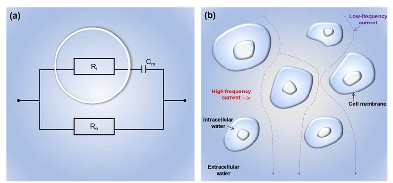
Frequency-dependent electrical behavior of biological tissues and their equivalent circuit representation. (a) Cole model of biological tissue, in which Re, Ri an d Cm represent the extracellular resistance, intracellular resistance, and cell membrane capacitance, respectively. (b) Schematic illustration of frequency-dependent current flow through biological tissue. At low frequencies, the high reactance of the cell membrane restricts current flow primarily to the extracellular space. At high frequencies, the reduced membrane reactance allows current to pass through the cell membrane. Adapted from Ref. [25].
As illustrated in Fig. 1(b), at low frequencies, the high capacitive reactance of the cell membrane restricts current penetration into the intracellular compartment, causing current to flow predominantly through the extracellular fluid. Accordingly, low-frequency impedance primarily reflects extracellular fluid conditions and can be used to assess physiological changes associated with edema, dehydration, and fluid overload. In contrast, as the frequency increases, the capacitive reactance of the cell membrane decreases, allowing current to penetrate the cell membrane and flow through both the extracellular and intracellular compartments. High-frequency impedance therefore provides information on both extracellular and intracellular fluids and can be used to estimate body cell mass and nutritional status [25]. Thus, the physiological information reflected by bioimpedance measurements varies depending on the measurement frequency. Consequently, an appropriate frequency should be selected based on the target tissue and the specific parameter to be assessed.
In addition to measurement frequency, the accuracy and reliability of bioimpedance measurements are influenced by the electrode configuration and measurement technique. In a two-electrode configuration, the same pair of electrodes is used to inject current and measure the resulting voltage. In contrast, a four-electrode configuration separates the current-injecting electrodes from the voltage-sensing electrodes, thereby minimizing the influence of contact impedance on the measurement [28,29]. Bioimpedance techniques can also be classified according to the applied frequency range. Single-frequency bioelectrical impedance analysis (SFBIA) employs a single measurement frequency, whereas multiple-frequency bioelectrical impedance analysis (MFBIA) uses several discrete frequencies. Bioimpedance spectroscopy (BIS) measures impedance over a broad frequency range and fits the resulting spectrum to an appropriate electrical or biophysical model [6,25,27,30].
Recent advances in flexible electronic materials, miniaturized measurement circuits, and wireless communication technologies have enabled the development of wearable bioimpedance sensors. As a result, the application of bioimpedance technology has expanded beyond conventional body composition analysis to various physiological and clinical monitoring areas, including the cardiovascular, respiratory, and musculoskeletal systems [31-33].
3. WEARABLE BIOIMPEDANCE SENSORS
3.1 Muscle and body fluid monitoring
The biological components and tissues that make up the human body, such as body fluids, muscle, and fat, each have distinct electrical conductivity and permittivity. Therefore, by tracking changes in measured bioimpedance, subtle physiological and structural changes within the body can be quantified [34]. Fig. 2 shows a representative example of a bioimpedance-based wearable device for muscle monitoring [28]. The flexible multichannel electrical impedance sensor (FMEIS) was designed to overcome the limitations of EMG, a conventional wearable interface, and Fig. 2(a) illustrates the fundamental operating mechanism of FMEIS. FMEIS delivers a low-amplitude, high-frequency alternating current into subcutaneous tissue without inducing any perceptible sensation in the human body. Such high-frequency, low-amplitude excitation is suitable for muscle impedance measurements because it enables the impedance response to be monitored without intentionally eliciting sensory or muscular stimulation [28]. When a muscle contracts or changes shape, the internal electrical conductivity distribution changes, causing distortion in the electric field. The multichannel silver circuits on the sensor surface capture these subtle voltage changes to reconstruct muscle movement accurately. For such non-invasive impedance measurements to be possible, the sensor must conform closely to the skin surface. Fig. 2(b) shows the ultrathin, flexible structure of the FMEIS designed to achieve this conformal contact. This sensor consists of a polydimethylsiloxane (PDMS) encapsulation layer surface-modified with ethoxylated polyethyleneimine (PEIE), a conductive silver circuit, and a hydrogel electrode that comes into direct contact with the skin. With a total thickness of only 220 μm and a low elastic modulus similar to that of human skin, the sensor adheres firmly to the skin via hydrogen bonding, thereby minimizing motion artifacts that may occur during movement. Fig. 2(c) demonstrates the stability of biological signals extracted using this skin-adhering design. When the sensor was attached to a subject’s forearm and the flexor carpi radialis (FCR) and extensor carpi radialis (ECR) muscles were monitored using a four-point impedance measurement method, distinct and regular impedance waveforms were measured during repeated wrist extension bending the wrist upward and flexion (bending the wrist downward) movements. Consistent with the results of the cross-sectional electric field distribution simulation, localized impedance changes were precisely captured in response to the activation of specific muscles. Finally, Fig. 2(d) provides a comparative analysis of the physiological advantages that FMEIS technology offers over conventional EMG sensors. In the graph at the bottom of Fig. 2(d)(ii), commercial EMG sensors detect electrical activity signals only when the FCR or ECR muscles are actively contracting, revealing a fundamental limitation in that they fail to capture signals when the opposing muscles are passively relaxed [35]. In contrast, compared with conventional EMG, which primarily detects the electrical activity of actively contracting muscles, FMEIS can capture both active contraction and passive muscle deformation [28]. In conclusion, owing to its flexible and biocompatible form factor (Figs. 2(a) and (b)), FMEIS achieves stable signal acquisition even during dynamic movement (Fig. 2(c)). By successfully capturing passive muscle deformation states that conventional technologies miss (Fig. 2(d)), this system offers an effective muscle force prediction solution for next-generation human-machine interfaces (HMIs). Such technology, which accurately reconstructs muscle activity without delay, could enable future HMIs to provide an environment that is fully integrated with the human body [36]. To this end, research is currently underway to combine FMEIS with multimodal integration technology that simultaneously captures tactile and bioelectric signals, as well as ion-based, biocompatible sensing technology that mimics the body’s natural signal transmission mechanisms [37,38].
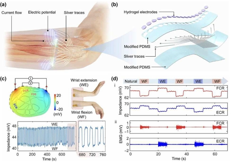
A muscle activity monitoring system based on a FMEIS. (a) Operating mechanism of FMEIS. (b) Structure of the ultrathin, flexible sensor designed for perfect skin contact. (c) Stable impedance waveforms captured on the forearm during wrist flexion and extension, along with simulation results of the local electric field distribution. (d) Performance comparison between FMEIS (i), which captures even the deformation associated with passive muscle relaxation, and existing commercial EMG sensors (ii), which detect only active muscle contractions. Adapted from Ref. [28].
Recently, wearable bioimpedance technology has moved beyond simple static body composition analysis to offer significant advantages in noninvasively and continuously tracking dynamic fluid changes within the body [39,40]. It is also being expanded into various clinical applications, such as fluid monitoring for patients undergoing kidney dialysis, tracking breast milk intake in infants, and monitoring bladder volume [9,11,41,42]. These applications, including renal dialysis, breastfeeding, and bladder volume monitoring, all use the common principle that electrical impedance changes in response to increases or decreases in tissue fluid. For patients undergoing renal dialysis, multifrequency analysis is used to separate the resistance of intracellular and extracellular fluids, thereby quantifying fluid status in real time to prevent dehydration or fluid overload [41]. Furthermore, during breastfeeding, wearable bioimpedance systems can detect impedance changes that increase linearly with milk ejection, enabling continuous tracking of infant intake without the need for conventional pre- and post-feeding weight measurements [42]. Similarly, patients with urinary dysfunction can receive smartphone alerts indicating the optimal time to urinate by continuously measuring the decrease in impedance as urine fills the bladder [9,11].
3.2 Cardiovascular monitoring
Cyclic changes in arterial cross-sectional area and blood volume during the cardiac cycle can be captured as pulsatile bioimpedance waveforms [33,43,44]. The time delay between pulse waves recorded at two spatially separated arterial sites is defined as pulse transit time (PTT), and pulse wave velocity (PWV) can be calculated by dividing the distance between the two sites by the PTT [44-46]. As BP increases, arterial compliance generally decreases, and the pressure wave propagates more rapidly [47]. Accordingly, PWV increases, whereas PTT decreases [44,46,47]. However, the relationship between PTT and BP is affected by the measurement path length, arterial geometry, and other subject-specific factors and therefore generally requires individual calibration [43,47]. Compared with conventional cuff-based BP measurements, wearable bioimpedance offers the potential for continuous cuffless BP monitoring without repeated cuff inflation [19,33].
To translate these principles into wearable systems, electrode geometry and the stability of the skin interface must be carefully controlled [19,33,43,45]. Representative approaches include transverse dual-channel wrist configurations for PTT and PWV estimation [44], automatic gain control (AGC) circuits that compensate for variations in electrode–skin impedance [45], and compliant nanocomposite electrodes formed by combining Poly(3,4-ethylenedioxythiophene):poly(styrenesulfonate) (PEDOT:PSS) with silver nanowires (AgNWs) to improve signal quality [13,48]. Ring-shaped sensors provide another compact form factor because a properly sized ring can maintain repeatable contact over the digital arteries [19]. In addition to PTT-based methods, the morphology of a single-site pulsatile bioimpedance waveform can be analyzed using machine-learning models to estimate beat-to-beat BP [19].
Fig. 3 presents a four-electrode bioimpedance ring designed to capture pulsatile blood volume changes in the digital arteries [19]. The custom-sized ring maintains firm electrode-skin contact at a repeatable finger location (Fig. 3(a)). Because the limited ring area prevents conventional in-line placement of all four electrodes along the artery, a finite element model (FEM) incorporating the major finger tissues and digital arteries was used to analyze current distribution and optimize electrode placement (Fig. 3(b)). The device uses four 3 × 3 mm silver dry electrodes: one pair injects a 10 kHz alternating current, and the other pair measures the voltage response (Fig. 3(c)). This operating frequency provides sensitivity to impedance changes associated with pulsatile arterial blood flow and volume [19,46]. Cardiac-cycle-dependent expansion of the digital artery increases local blood volume and reduces the measured impedance during systole, whereas impedance increases as blood volume leaves the sensing region during diastole. Amplitude, timing, area, and derivative features extracted from each bioimpedance pulse were subsequently used in AdaBoost regression models to estimate systolic BP (SBP) and diastolic BP (DBP). In 10 healthy male participants, BP variations were induced using repeated cold pressor tests and rest periods, resulting in SBP and DBP ranges of 89–213 and 42–122 mmHg, respectively. Within these ranges, the system achieved mean errors of 0.11 ± 5.27 mmHg for SBP and 0.11 ± 3.87 mmHg for DBP, with average Pearson correlation coefficients of 0.76 and 0.81, respectively. These results demonstrate the potential of a compact ring form factor for continuous cuffless BP monitoring. Nevertheless, the models were trained and validated mainly using participant-specific data from a small cohort of young, healthy adults. Therefore, larger studies are required to establish interindividual generalizability, long-term stability, and performance during daily activities [19].
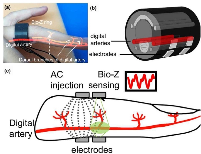
Bioimpedance ring sensor for arterial blood-flow and pressure monitoring. (a) Custom-fitted ring positioned over the digital arteries. (b) Finite element model of the finger used to guide the ring design and electrode placement. (c) Cross-sectional illustration of four-electrode bioimpedance sensing, showing high-frequency AC distribution and the region sensitive to arterial volume changes. Adapted from Ref. [19].
3.3 Respiratory monitoring
Respiratory rate (RR) is a key vital sign that may provide early indications of hypoxia, hypercapnia, and overall physiological deterioration [49]. However, intermittent manual measurement by clinical staff may be missed or inaccurate, and awareness of being observed can alter a patient's spontaneous breathing pattern. Therefore, noninvasive technologies capable of continuously monitoring respiration without disrupting daily activities are needed [50-52].
Impedance pneumography (IP) is a noninvasive respiratory-monitoring method that measures respiration-induced changes in thoracic bioimpedance [50,53]. As pulmonary air volume and thoracic geometry change during breathing, thoracic bioimpedance varies cyclically, increasing during inspiration and decreasing during expiration [14,53]. These respiratory impedance changes reflect not only changes in lung-tissue resistivity as the lungs fill with air but also changes in thoracic circumference, interelectrode distance, diaphragmatic position, and the resulting current pathways [53]. IP can be implemented using two- or four-electrode configurations. In the four-electrode configuration, the current-injection and voltage-sensing electrodes are separated, which reduces the effects of electrode-skin impedance and electrode motion [53].
RR and inspiratory and expiratory times can be derived from the periodicity and respiratory-phase boundaries of the bioimpedance waveform, whereas tidal volume (TV) can be estimated from its peak-to-peak amplitude [16,17,50]. Because the impedance-volume relationship varies among individuals and with posture and electrode placement, calibration against a reference instrument is required [17,50,54]. Seppä et al. evaluated IP in 20 healthy male participants across different TV levels and postures and reported good agreement between IP-derived flow waveforms and pneumotachograph reference waveforms. After subject- and posture-specific calibration, the difference in TV was less than 3% [54]. Thoracic bioimpedance reflects both lung-volume and chest-wall-motion components, with the contribution of chest-wall motion increasing under higher inspiratory loads [31]. Therefore, thoracic bioimpedance should be interpreted as a surrogate signal that jointly reflects pulmonary air-volume changes and thoracic geometry rather than as a direct measurement of lung volume [31,53]. Compared with intermittent manual RR assessment, wearable IP enables unobtrusive and continuous respiratory monitoring during daily activities [50-52]. However, TV is estimated indirectly from the impedance waveform and requires calibration against a reference measurement [17,50,54].
Conventional IP systems require widely spaced electrodes and external instrumentation, which limits wearability and signal stability during movement [50,53]. Miniaturization of the electrode array and measurement electronics has subsequently enabled sternum-mounted IP patches [50]. Berkebile et al. used a 5.1 × 5.1 cm tetrapolar patch to estimate RR and TV not only during sitting and supine rest but also during walking and stair climbing. The estimated TV was correlated with measurements from a reference spirometer [50].
Recent wearable respiratory platforms integrate flexible or stretchable sensing elements, miniaturized signal-processing electronics, and wireless communication to collect physiological signals continuously during daily activities [55]. Rather than completely replacing conventional pulmonary function tests, these devices may serve as adjunctive tools for tracking respiratory changes between clinical visits and identifying abnormal breathing patterns at an early stage [52]. Sanchez-Perez et al. integrated multifrequency IP, BIS, an inertial sensor, and multichannel lung-sound sensors in hospitalized patients with heart failure, enabling the extraction of RR and the expiratory-to-inspiratory time ratio and the identification of Cheyne-Stokes respiration [17].
Qiu et al. developed a rigid-flex chest patch integrating bioimpedance, an inertial measurement unit (IMU), Bluetooth, and long-range (LoRa) wireless communication for real-time RR monitoring. The system achieved accuracy above 97.8% under static conditions and above 98.5% during walking, running, and cycling [14]. Together, these studies show that IP-based bioimpedance can provide RR, respiratory timing, and TV estimates, while multimodal systems such as seismocardiography, photoplethysmography, impedance pneumography and cardiography, surface respiratory mechanomyography and electromyography, and electrocardiography can additionally assess the mechanical and electrical activity of the respiratory muscles [14,16,17,50].
Fig. 4 summarizes IP-based respiratory monitoring and TV estimation using the chest-worn reSPIRE system [16]. As shown in Fig. 4(a), the device is positioned symmetrically about the sternum, with two sensing arms placed over the second intercostal spaces adjacent to the sternum. Four IP electrodes are arranged from left to right as the negative current-injection electrode (I-), negative voltage-sensing electrode (V-), positive voltage-sensing electrode (V+), and positive current-injection electrode (I+). Representative IP waveforms acquired during spontaneous breathing and controlled variations in breathing rate and depth are shown in Fig. 4(b). Deep breaths produce larger IP amplitudes than shallow breaths, whereas the slow- and fast-breathing conditions correspond to approximately 6 and 15 breaths/min, respectively.
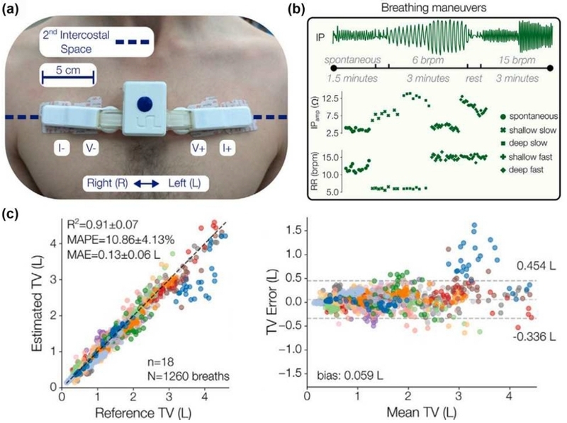
Chest-worn reSPIRE system for IP-based respiratory monitoring. (a) Device placement and tetrapolar IP electrode configuration. (b) Representative IP waveforms during spontaneous breathing and controlled changes in breathing rate and depth. (c) Comparison of IP-derived and spirometer-measured TV and corresponding Bland-Altman analysis. Adapted from Ref. [16].
As shown in Fig. 4(c), IP-based TV estimates agreed well with reference spirometer measurements, with R² = 0.91 ± 0.07, a mean absolute error (MAE) of 0.135 ± 0.063 L, and a mean absolute percentage error (MAPE) of 10.86 ± 4.13%. Bland-Altman analysis yielded a mean bias of 0.059 L and 95% limits of agreement from -0.336 to 0.454 L. These results demonstrate that IP-based bioimpedance can estimate RR, breathing depth, and TV, while surface respiratory mechanomyography (sRMG) and surface electromyography (sEMG) can assess the mechanical and electrical activity of the respiratory muscles, respectively. This multimodal information can help distinguish rapid, shallow breathing from slow, deep breathing patterns that may not be differentiated by RR alone, supporting potential applications in chronic obstructive pulmonary disease (COPD) and disorders involving respiratory-muscle weakness [15,16,51]. However, IP is sensitive to motion, posture, electrode position, and electrode-skin contact and may overlap with cardiogenic artifacts. Therefore, subject-specific calibration remains necessary for TV estimation [50,51,53,54]. Furthermore, reSPIRE was validated only in 18 healthy participants, highlighting the need for standardized, long-term studies in diverse patient populations and real-world settings [16,51,52,54]. Therefore, quantitative performance reported across these studies should be interpreted within the context of their respective participant populations, experimental protocols, calibration procedures, and reference methods rather than directly compared.
3.4 Skin and tissue monitoring
Conventional diagnosis of skin and tissue disorders has largely relied on visual inspection and optical assessment, which may not sufficiently reflect physiological information from within the tissue [56,57]. In addition, these approaches can be influenced by the subjective judgment of clinicians and have limitations for repeated monitoring, including an increased risk of infection associated with dressing removal [58]. Impedance-based sensors provide a nonvisual diagnostic approach and have therefore emerged as an alternative approach for addressing these limitations [22]. The electrical properties of biological tissues reflect their physiological state, and changes in the skin are accompanied by corresponding changes in bioimpedance. Accordingly, wearable bioimpedance sensors have been actively developed to enable real-time monitoring and provide objective, quantitative information on skin and tissue conditions [59,60]. Compared with visual and optical assessment alone, bioimpedance can provide objective, quantitative information on skin and tissue conditions and may support repeated assessment without frequent dressing removal [56-58]. In general, damaged skin and tissues exhibit lower impedance than healthy tissues. This difference can primarily be attributed to cell membrane damage and changes in the local tissue environment. Skin and tissue injury is accompanied by cell loss and disruption of cell membrane integrity, whereas the accumulation of ion-rich exudate increases tissue conductivity and consequently reduces impedance [61]. Keratinocytes, which constitute the major cellular component of the epidermis, also play an important role in determining skin bioimpedance [62]. In skin tissue exposed to inflammatory cytokines, reduced expression of keratinocyte differentiation markers impairs skin barrier function, thereby decreasing bioimpedance. During the subsequent wound-healing process, epidermal proliferation and granulation tissue formation restore tissue structure, resulting in a gradual increase in impedance toward the level of healthy skin [63].
Fig. 5(a) presents an overview of a wearable system for measuring the bioimpedance of skin and underlying tissues. The reader module wirelessly transfers power to the patch through magnetic induction, and the powered patch applies an electrical current to the tissue through its electrodes. These electrodes are in direct contact with the skin, enabling measurement of tissue bioimpedance. Changes in tissue bioimpedance alter the resonant characteristics of the system, which are detected by the reader module to infer the electrical properties of the tissue. Figs. 5(b) and (c) compare the resistance and capacitance of skin lesions represented by moles with those of healthy skin, demonstrating lower resistance and higher capacitance in the moles. These differences in the resistive and capacitive components provide an electrical basis for distinguishing the lesions from healthy skin based on bioimpedance [56]. In addition, because the penetration depth of the applied current and the tissue region contributing to the measured signal vary with frequency, the measurement frequency must be selected according to the intended application. Based on these frequency-dependent characteristics, bioimpedance-based monitoring has recently expanded beyond localized skin lesions to a broader range of skin and tissue disorders, including chronic wounds, diabetic foot ulcers, and burns. Furthermore, these systems are evolving into closed-loop platforms capable of simultaneous monitoring and treatment, and their integration with machine-learning models may enable the development of personalized skin and tissue management systems [10,22].
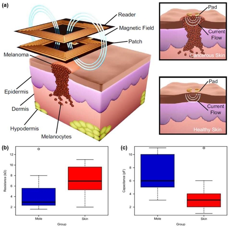
Wireless wearable bioimpedance sensing system for clinical skin evaluation. (a) Schematic illustration of a wearable bioimpedance sensing system in which the reader wirelessly couples with the skin-mounted patch through magnetic induction. (b), (c) Comparison of the resistance and capacitance measured in moles and healthy skin, respectively. Adapted from Ref. [56].
4. CONCLUSIONS
With the aging of society and the increasing prevalence of chronic diseases, the importance of wearable bioimpedance technology, which can monitor physiological changes at the cellular and tissue levels, is increasing. As the medical paradigm shifts from post-disease treatment to a focus on daily prevention and management, the preventive medical value and socioeconomic benefits of this technology are receiving even greater attention. Furthermore, with recent advancements in flexible electronic materials and multi-electrode measurement circuits, the scope of bioimpedance technology is expanding beyond body composition analysis to a wide range of areas, including the cardiovascular and respiratory systems, muscles, body fluids, and skin lesions. These systems share the common advantage of monitoring subtle electrical changes in tissues noninvasively and continuously in everyday environments. Specifically, they have demonstrated the potential to quantitatively assess muscle contraction and relaxation states, cardiovascular indicators for cuffless blood pressure measurement, respiratory rate and tidal volume, as well as skin lesions and healing status.
However, several limitations must still be addressed for the clinical application of wearable bioimpedance technology. Although bioimpedance signals reflect the physiological characteristics of tissues, they are also influenced by an individual’s anatomical characteristics, posture, electrode placement, and skin-electrode contact conditions. Consequently, impedance values obtained from different users and measurement environments are difficult to use as common reference values, and similar impedance changes may result from different causes. During prolonged wear, movement, electrode displacement, contact-pressure variation, and changes in skin condition can cause signal fluctuations that are difficult to distinguish from actual physiological changes. Over long-term and real-world use, these effects may accumulate as baseline drift, and their magnitude can vary among users because of differences in anatomy, tissue composition, skin condition, and daily activity patterns. Furthermore, issues such as optimizing power consumption, improving form factors, and ensuring the security and privacy protection of the vast amounts of sensitive personal biometric data collected during long-term continuous measurement are also challenges that must be addressed for commercialization.
To overcome these limitations, future research on wearable bioimpedance should focus not only on improving sensor sensitivity but also on enhancing measurement reliability and personalizing signal interpretation. To this end, it is necessary to standardize measurement conditions, such as electrode placement and measurement frequency, while individual baselines and periodic signal-quality checks should be used to track trends over time. Moreover, measurement specificity must be improved by distinguishing changes caused by movement and wearing conditions from actual physiological changes through multimodal integration with biometric data, such as body temperature, perspiration, and optical signals. Building on these advances, artificial intelligence and cloud computing technologies can be leveraged to train systems using large-scale real-world usage data, enabling the early detection of disease signs based on individual baselines and the accurate prediction of condition deterioration. Crucially, medical device approval through rigorous clinical validation will be essential for this technology to move beyond simple wellness devices and be integrated into actual clinical settings.
Ultimately, by expanding into a closed-loop system that links the detection of abnormal conditions to therapeutic processes such as electrical stimulation and drug delivery, this technology could evolve into a core technology for implementing a personalized treatment platform in which disease diagnosis and treatment are closely integrated. This development could play an important role in extending healthy life expectancy through continuous monitoring in daily life within the emerging digital healthcare ecosystem.
Acknowledgments
This work was supported by the National Research Foundation of Korea (NRF) grant funded by the Korea government (MSIT) (RS-2026-25499347). This work was supported by the National Research Foundation of Korea (NRF) grant funded by the Korea government (MSIT) (RS-2025-24202976). This research was supported by Basic Science Research Program through the National Research Foundation of Korea (NRF) funded by the Ministry of Education (RS-2025-25432139). This work was supported by the Ajou University research fund. This research was supported by Global - Learning & Academic research institution for Master’s·PhD students, and Postdocs (G-LAMP) Program of the National Research Foundation of Korea (NRF) grant funded by the Ministry of Education (No. RS-2023-00285390).
REFERENCES
-
E. Borelli, G. Paolini, F. Antoniazzi, M. Barbiroli, F. Benassi, F. Chesani, et al., HABITAT: An IoT solution for independent elderly, Sensors 19 (2019) 1258.
[https://doi.org/10.3390/s19051258]

-
D. Naranjo-Hernández, J. Reina-Tosina, M. Min, Fundamentals, recent advances, and future challenges in bioimpedance devices for healthcare applications, J. Sens. 2019 (2019) 9210258.
[https://doi.org/10.1155/2019/9210258]
-
M. Lin, H. Hu, S. Zhou, S. Xu, Soft wearable devices for deep-tissue sensing, Nat. Rev. Mater. 7 (2022) 850–869.
[https://doi.org/10.1038/s41578-022-00427-y]
-
N. Arabsalmani, A. Ghouchani, S. Jafarabadi Ashtiani, M. Zamani, Exploring bio-impedance sensing for intelligent wearable devices, Bioengineering 12 (2025) 521.
[https://doi.org/10.3390/bioengineering12050521]
-
R. He, H. Liu , Y. Niu , H. Zhang, G.M. Genin, F. Xu , Flexible miniaturized sensor technologies for long-term physiological monitoring, npj Flex. Electron. 6 (2022) 20.
[https://doi.org/10.1038/s41528-022-00146-y]
-
A.W. Potter, L.C. Ward, C.L. Chapman, W.J. Tharion, D.P. Looney, K.E. Friedl, Real-world assessment of multi-frequency bioelectrical impedance analysis (MFBIA) for measuring body composition in healthy physically active populations, Eur. J. Clin. Nutr. 79 (2025) 1235–1244.
[https://doi.org/10.1038/s41430-025-01664-4]
-
J.H. Kim, S.S. Kim, S.H. Kim, S.W. Baik, G.R. Jeon, Bioelectrical impedance analysis at popliteal regions of human body using BIMS, J. Sens. Sci. Technol. 25 (2016) 1–7.
[https://doi.org/10.5369/JSST.2016.25.1.1]
-
C.F. Brandner, G.M. Tinsley, A.J. Graybeal, Smartwatch-based bioimpedance analysis for body composition estimation: Precision and agreement with a 4-compartment model, Appl. Physiol. Nutr. Metab. 48 (2023) 172–182.
[https://doi.org/10.1139/apnm-2022-0301]
-
S. Lim, J. Kim, Wireless bioelectronics for bladder function monitoring, J. Sens. Sci. Technol. 34 (2025) 513–519.
[https://doi.org/10.46670/JSST.2025.34.5.513]
-
B. Li, Y. Wang, J. Mei, X. He, Wearable bioelectronics: Toward future personal health monitoring, BIO Integr. 7 (2026) 1–10.
[https://doi.org/10.15212/bioi-2025-0219]
-
L. Duan, M.L. Jin, A biocompatible integrated bladder electronics for wireless capacity monitoring assessment, Soft Sci. 5 (2025) 4.
[https://doi.org/10.20517/ss.2024.46]
-
A. Mokaddem, D. Ziani, M. Belkheir, M. Rouissat, B. Doumi, P. Lorenz, Eco-Friendly Circular Meandered Loop Antenna Design Based on Novel PLA/PHBV Biocomposite and SWCNT Conductive Material for Medical Applications, Trans. Electr. Electron. Mater. 26 (2025) 931–944.
[https://doi.org/10.1007/s42341-025-00637-5]
-
M. Namkoong, B. Baskar, L. Singh, H. Guo, J. McMurray, K. Branan, et al., Add-on soft electronic interfaces for continuous cuffless blood pressure monitoring, Adv. Mater. Technol. 8 (2023) 2300158.
[https://doi.org/10.1002/admt.202300158]
-
C. Qiu , F. Wu , W. Han, M.R. Yuce, A wearable bioimpedance chest patch for real-time ambulatory respiratory monitoring, IEEE Trans. Biomed. Eng. 69 (2022) 2970–2981.
[https://doi.org/10.1109/TBME.2022.3158544]
-
D. Blanco-Almazán, W. Groenendaal, M. Lozano-García, L. Estrada-Petrocelli, L. Lijnen, C. Smeets, et al., Combining bioimpedance and myographic signals for the assessment of COPD during loaded breathing, IEEE Trans. Biomed. Eng. 68 (2021) 298–307.
[https://doi.org/10.1109/TBME.2020.2998009]
-
J.A. Berkebile, H.T. Crane, J.A. Sánchez-Pérez, K. Swamy, F.N. Rahman, O.T. Inan, reSPIRE: A wearable multimodal sensing system for monitoring ventilation, cardiovascular dynamics, and respiratory muscle activity, Biosens. Bioelectron. 301 (2026) 118460.
[https://doi.org/10.1016/j.bios.2026.118460]
-
J.A. Sanchez-Perez, J.A. Berkebile, B.N. Nevius, G.C. Ozmen, C.J. Nichols, V.G. Ganti, et al., A wearable multimodal sensing system for tracking changes in pulmonary fluid status, lung sounds, and respiratory markers, Sensors 22 (2022) 1130.
[https://doi.org/10.3390/s22031130]
-
Z. Ghemari, A. Bendaikha, S. Belkhiri, S. Saad, Fabrication of Flexible Resistive Pressure Sensors Using Graphene/Polydimethylsiloxane Composites, Trans. Electr. Electron. Mater. 26 (2025) 323–344.
[https://doi.org/10.1007/s42341-025-00601-3]
-
K. Sel, D. Osman, N. Huerta, A. Edgar, R.I. Pettigrew, R. Jafari, Continuous cuffless blood pressure monitoring with a wearable ring bioimpedance device, npj Digit. Med. 6 (2023) 59.
[https://doi.org/10.1038/s41746-023-00796-w]
-
H.C. Ates, P.Q. Nguyen, L. Gonzalez-Macia, E. Morales-Narváez, F. Güder, J.J. Collins, et al., End-to-end design of wearable sensors, Nat. Rev. Mater. 7 (2022) 887–907.
[https://doi.org/10.1038/s41578-022-00460-x]
-
M. Shwetha, S. Lakshmi, Harmless Sustainable Energy Resource using TEG with MPPT Based DC-DC Converter for Power Gain in Implantable Medical Devices, Trans. Electr. Electron. Mater. 25 (2024) 608–635.
[https://doi.org/10.1007/s42341-024-00539-y]
-
Y. Jiang, A.A. Trotsyuk, S. Niu, D. Henn, K. Chen, C.-C. Shih, et al., Wireless, closed-loop, smart bandage with integrated sensors and stimulators for advanced wound care and accelerated healing, Nat. Biotechnol. 41 (2023) 652–662.
[https://doi.org/10.1038/s41587-022-01528-3]
-
T.K. Bera, Bioelectrical impedance methods for noninvasive health monitoring: A review, J. Med. Eng. 2014 (2014) 381251.
[https://doi.org/10.1155/2014/381251]
-
I. Jacobs, A. Lowe, L. Garcia, H. Zhang, Advancing physical activity monitoring through bioimpedance measurement: A review, Prog. Biomed. Eng. 8 (2026) 022002.
[https://doi.org/10.1088/2516-1091/ae3671]
-
J.R. Matthie, Bioimpedance measurements of human body composition: Critical analysis and outlook, Expert Rev. Med. Devices 5 (2008) 239–261.
[https://doi.org/10.1586/17434440.5.2.239]
-
L.C. Ward, S. Brantlov, Bioimpedance basics and phase angle fundamentals, Rev. Endocr. Metab. Disord. 24 (2023) 381–391.
[https://doi.org/10.1007/s11154-022-09780-3]
-
S. Brantlov, L.C. Ward, S. Isidor, C.L. Hvas, C.L. Rud, L. Jødal, Cell membrane capacitance (Cm) measured by bioimpedance spectroscopy (BIS): A narrative review of its clinical relevance and biomarker potential, Sensors 25 (2025) 4362.
[https://doi.org/10.3390/s25144362]
-
J. Li, K. Wu, J. Xiao, T. Chen, X. Yang, J. Pan, et al., Flexible multichannel muscle impedance sensors for collaborative human-machine interfaces, Sci. Adv. 11 (2025) eadv3359.
[https://doi.org/10.1126/sciadv.adv3359]
-
C. Bester, T. Razmovski, A. Collins, O. Mejia, S. Foghsgaard, A. Mitchell-Innes, et al., Four-point impedance as a biomarker for bleeding during cochlear implantation, Sci. Rep. 10 (2020) 2777.
[https://doi.org/10.1038/s41598-019-56253-w]
-
C.M. Prado, M.C. Gonzalez, K. Norman, C.E. Orsso, R. Barazzoni, T. Cederholm, et al., Methodological standards for body composition assessment—an expert-endorsed guide for research and clinical applications: bioimpedance, dual-energy X-ray absorptiometry, computerized tomography, and ultrasound methods, Am. J. Clin. Nutr. 123 (2026) 101283.
[https://doi.org/10.1016/j.ajcnut.2026.101283]
-
D. Blanco-Almazán, W. Groenendaal, F. Catthoor, R. Jané, Chest movement and respiratory volume both contribute to thoracic bioimpedance during loaded breathing, Sci. Rep. 9 (2019) 20232.
[https://doi.org/10.1038/s41598-019-56588-4]
-
E.A. Bondareva, O.I. Parfenteva, E.A. Troshina, E.V. Ershova, N.V. Mazurina, K.A. Komshilova, et al., Agreement between bioimpedance analysis and ultrasound scanning in body composition assessment, Am. J. Hum. Biol. 36 (2024) e24001.
[https://doi.org/10.1002/ajhb.24001]
-
D. Kireev, K. Sel, B. Ibrahim, N. Kumar, A. Akbari, R. Jafari, et al., Continuous cuffless monitoring of arterial blood pressure via graphene bioimpedance tattoos, Nat. Nanotechnol. 17 (2022) 864–870.
[https://doi.org/10.1038/s41565-022-01145-w]
-
Y.I. Shin, G.H. Kim, Y.J. Hwang, S.W. Baik, J.H. Kim, G.R. Jeon, Assessment of body fluid alteration using bioelectrical impedance in stroke patients with hemiplegia caused by cerebral hemorrhage and cerebral infarction, J. Sens. Sci. Technol. 26 (2017) 160–167.
[https://doi.org/10.5369/JSST.2017.26.3.160]
- M.L. Latash, Fundamentals of Motor Control, first ed., Academic Press, San Diego, 2012.
-
K.K. Kim, T.J. Zaluska, S. Skov, Y. Lee, H. Park, D. Zhong, et al., A simplified wearable device powered by a generative EMG network for hand-gesture recognition and gait prediction, Nat. Sens. 1 (2026) 27–38.
[https://doi.org/10.1038/s44460-025-00002-2]
-
P. Wan, D. Yao, S. Wan, S. Li, Y. Ye, H. Huang, et al., A bioinspired ionic diode tactile sensor for embodied sensorimotor intelligence in robotics, Adv. Funct. Mater. 36 (2026) e76889.
[https://doi.org/10.1002/adfm.76889]
-
Z. Wang, W. Yang, X. Wang, D. Xing, S. Liu, Y. Wang, et al., Flexible bimodal sensor with integrated tactile and bioelectrical signal perception for human-computer interaction systems, Adv. Funct. Mater. 36 (2026) e75491.
[https://doi.org/10.1002/adfm.75491]
-
M.H. Jung, K. Namkoong, Y. Lee, Y.J. Koh, K. Eom, H. Jang, et al., Wrist-wearable bioelectrical impedance analyzer with miniature electrodes for daily obesity management, Sci. Rep. 11 (2021) 1238.
[https://doi.org/10.1038/s41598-020-79667-3]
-
D.M. Davydov, A. Boev, S. Gorbunov, Making the choice between bioelectrical impedance measures for body hydration status assessment, Sci. Rep. 11 (2021) 7685.
[https://doi.org/10.1038/s41598-021-87253-4]
-
D. Allegri, D. Vaca, D. Ferreira, M. Rogantini, D. Barrettino, Real-time monitoring of the hydration level by multi-frequency bioimpedance spectroscopy, Proceedings of the 2017 IEEE International Instrumentation and Measurement Technology Conference (I2MTC), Turin, Italy, 2017, pp. 1–6.
[https://doi.org/10.1109/I2MTC.2017.7969668]
-
J. Kim, S. Oh, R. Avila, H.-S. Shin, M. Banet, J. Wicks, et al., A compact, wireless system for continuous monitoring of breast milk expressed during breastfeeding, Nat. Biomed. Eng. 9 (2025) 1645–1655.
[https://doi.org/10.1038/s41551-025-01393-w]
-
G. Anand, Y. Yu, A. Lowe, A. Kalra, Bioimpedance analysis as a tool for hemodynamic monitoring: Overview, methods and challenges, Physiol. Meas. 42 (2021) 03TR01.
[https://doi.org/10.1088/1361-6579/abe80e]
-
T.H. Huynh, R. Jafari, W.-Y. Chung, An accurate bioimpedance measurement system for blood pressure monitoring, Sensors 18 (2018) 2095.
[https://doi.org/10.3390/s18072095]
-
B. Passage, B. Ibrahim, R. Jafari, Real-time signal-to-noise ratio optimization of bio-impedance signal for cuffless blood pressure monitoring, Proceedings of the 43rd Annual International Conference of the IEEE Engineering in Medicine and Biology Society (EMBC), Virtual Conference, 2021, pp. 7480–7484.
[https://doi.org/10.1109/EMBC46164.2021.9630920]
-
M. Namkoong, J. McMurray, K. Branan, J. Hernandez, M. Gandhi, S. Ida-Oze, et al., Contact pressure-guided wearable dual-channel bioimpedance device for continuous hemodynamic monitoring, Adv. Mater. Technol. 9 (2024) 2301407.
[https://doi.org/10.1002/admt.202301407]
-
R. Mukkamala, J.-O. Hahn, O.T. Inan, L.K. Mestha, C.-S. Kim, H. Töreyin, et al., Toward ubiquitous blood pressure monitoring via pulse transit time: Theory and practice, IEEE Trans. Biomed. Eng. 62 (2015) 1879–1901.
[https://doi.org/10.1109/TBME.2015.2441951]
-
M. Namkoong, H. Guo, M.S. Rahman, D. Wang, C.J. Pfeil, S. Hager, et al., Moldable and transferrable conductive nanocomposites for epidermal electronics, npj Flex. Electron. 6 (2022) 41.
[https://doi.org/10.1038/s41528-022-00170-y]
-
S. Rolfe, The importance of respiratory rate monitoring, Br. J. Nurs. 28 (2019) 504–508.
[https://doi.org/10.12968/bjon.2019.28.8.504]
-
J.A. Berkebile, S.A. Mabrouk, V.G. Ganti, A.V. Srivatsa, J.A. Sanchez-Perez, O.T. Inan, Towards estimation of tidal volume and respiratory timings via wearable-patch-based impedance pneumography in ambulatory settings, IEEE Trans. Biomed. Eng. 69 (2022) 1909–1919.
[https://doi.org/10.1109/TBME.2021.3130540]
-
M. Folke, L. Cernerud, M. Ekström, B. Hök, Critical review of non-invasive respiratory monitoring in medical care, Med. Biol. Eng. Comput. 41 (2003) 377–383.
[https://doi.org/10.1007/BF02348078]
-
B.A. Vicente, R. Sebastião, V. Sencadas, Wearable devices for respiratory monitoring, Adv. Funct. Mater. 34 (2024) 2404348.
[https://doi.org/10.1002/adfm.202404348]
-
L.E. Baker, Applications of the impedance technique to the respiratory system, IEEE Eng. Med. Biol. Mag. 8 (1989) 50–52.
[https://doi.org/10.1109/51.32406]
-
V.-P. Seppä, J. Viik, J. Hyttinen, Assessment of pulmonary flow using impedance pneumography, IEEE Trans. Biomed. Eng. 57 (2010) 2277–2285.
[https://doi.org/10.1109/TBME.2010.2051668]
-
R.A. Shaukat, A.M. Tamim, G.T. Hwang, C.K. Jeong, Humidity Sensors Using 2D and 3D Nanomaterials: From Materials Selection to Technological Aspects, Trans. Electr. Electron. Mater. 25 (2024) 123–140.
[https://doi.org/10.1007/s42341-024-00508-5]
-
H. Imtiaz, E. Ray, E. Lim, M. Tahernezhadi, M.J. Moghimi, Wearable battery-free chip-less patch for bioimpedance measurement of cutaneous lesions, npj Biomed. Innov. 2 (2025) 32.
[https://doi.org/10.1038/s44385-025-00037-7]
- A. Kekonen, M. Bergelin, J.-E. Eriksson, H. Ylänen, J. Viik, A quantitative method for monitoring wound healing, Int. J. Bioelectromagn. 17 (2015) 36–41.
-
A. Kekonen, M. Bergelin, J.-E. Eriksson, A. Vaalasti, H. Ylänen, S. Kielosto, et al., Bioimpedance method for monitoring venous ulcers: Clinical proof-of-concept study, Biosens. Bioelectron. 178 (2021) 112974.
[https://doi.org/10.1016/j.bios.2021.112974]
-
P. Yang, Z. Liu, S. Qin, J. Hu, S. Yuan, Z.L. Wang, et al., A wearable triboelectric impedance tomography system for noninvasive and dynamic imaging of biological tissues, Sci. Adv. 10 (2024) eadr9139.
[https://doi.org/10.1126/sciadv.adr9139]
-
M.I. Hossain, M.S. Zahid, M.A. Chowdhury, M.M.M. Hossain, N. Hossain, M.A. Islam, et al., Smart bandage: A device for wound monitoring and targeted treatment, Results Chem. 7 (2024) 101292.
[https://doi.org/10.1016/j.rechem.2023.101292]
-
J.-H. Kim, B.-J. Shin, S.-W. Baik, G.-R. Jeon, Early detection of intravenous infiltration using multi-frequency bioelectrical impedance parameters: Pilot study, J. Sens. Sci. Technol. 26 (2017) 15–23.
[https://doi.org/10.5369/JSST.2017.26.1.15]
-
N.J.M. van den Brink, F. Pardow, L.D. Meesters, I. van Vlijmen-Willems, D. Rodijk-Olthuis, H. Niehues, et al., Electrical impedance spectroscopy quantifies skin barrier function in organotypic in vitro epidermis models, J. Invest. Dermatol. 144 (2024) 2488–2500.
[https://doi.org/10.1016/j.jid.2024.03.038]
-
L. Wang, G. Yao, S. Cai, X. Gan, Y. Gan, Y. Ren, et al., Modifiable PEDOT:PSS-based flexible diagnostic and therapeutic dressings for personalized wound management, Microsyst. Nanoeng. 12 (2026) 148.
[https://doi.org/10.1038/s41378-026-01200-9]

Kyungmin Kim is currently an M.S. student in the Department of Molecular Science and Technology at Ajou University. His research interests focus on wearable bioimpedance sensors for continuous health monitoring, particularly their applications in skin, tissue, and wound monitoring.

Dae A Wee is currently an M.S. student in the Department of Molecular Science and Technology at Ajou University. His research focuses on wearable bioimpedance sensors for physiological and muscle monitoring.

HyunA Jeong is an undergraduate student in the Department of Applied Chemistry & Biological Engineering at Ajou University. Her research focuses on wearable bioimpedance technology and non-invasive bioelectronics for continuous healthcare monitoring.

Jihye Kim is an Assistant Professor in the College of Advanced Bio-convergence Engineering and Department of Molecular Science and Technology at Ajou University. She received her B.S. and Ph.D. degrees in the School of Advanced Materials Science and Engineering from Sungkyunkwan University in Korea in 2015 and 2020, respectively. She then moved to Northwestern University as a postdoctoral scholar in the Center for Bio-Integrated Electronics (CBIE). Her research interests include wearable/implantable bioelectronics for physiological fluid monitoring. For more information, please visit the website: http://jkim.ajou.ac.kr.