
Recent Advances in Piezoelectric Ultrasound Sensors: A Compact Review on Key Applications

ⓒ The Korean Sensors Society
This is an Open Access article distributed under the terms of the Creative Commons Attribution Non-Commercial License (https://creativecommons.org/licenses/by-nc/3.0/) which permits unrestricted non-commercial use, distribution, and reproduction in any medium, provided the original work is properly cited.
Abstract
Piezoelectric ultrasound sensors are essential across biomedical monitoring, distance and obstacle detection, structural health monitoring, and emerging fields such as battery diagnostics and wireless energy transfer, owing to their high electromechanical coupling and fast response. This review provides a comprehensive overview of piezoelectric ultrasound sensing modules and their applications. We first examine the key material parameters that govern sensor performance, including the piezoelectric charge and voltage coefficients, the electromechanical coupling factor, and the mechanical quality factor, together with strategies such as phase-boundary engineering, dopant engineering, and texturing used to enhance them. It then surveys four major application domains such as bio-signal sensing, obstacle or distance sensing in different environmental conditions, and structural sensing by summarizing the key sensor design considerations that are relevant to each domain, along with representative studies. Finally, we discuss forward-looking strategies to overcome current performance limits, including acoustic metamaterial amplification, biocompatible and tissue-integrated material design, and lead-free piezoelectric development, outlining a path toward more sensitive, benign, and deeply integrated receiving modules in ultrasound sensors.
Keywords:
Piezoelectric ultrasound sensors, Biomedical sensors, Non-destructive testing, Structural health monitoring, Distance and obstacle detection, Battery diagnostics, Wireless energy transfer, Acoustic metamaterials, Dopant engineering, Templated grain growth, Acoustic impedance matching1. INTRODUCTION
Ultrasound-based sensors have become a common sensing modality in science and engineering [1]. They operate on the fundamental principle of propagating mechanical pressure waves and converting acoustic energy into measurable electrical signals [2]. While various source-based sensing technologies, such as optical, electromagnetic, and capacitive sensors, are widely used across modern industries, their penetration depth and resolution require improved performance for certain applications [3,4]. Ultrasound overcomes these limitations and demonstrates exceptional capabilities across several key application domains [5]. For example, in biomedical applications where ensuring human safety while securing reliable internal data is a major concern, ultrasound can be a suitable sensor source for maintaining high penetration as the source propagates through various internal tissues and operates without emitting harmful electromagnetic radiation [6-8]. Similarly, in ranging and obstacle detection sensors, it is crucial to transmit signals over a broad spatial range while minimizing severe scattering and erroneous signals [9]. Furthermore, structural failure detection also requires the sensing signal to successfully propagate through highly dense media [10]. In all these cases, ultrasound operates as an excellent source signal for sensors, inherently bypassing the fundamental limitations of other conventional sensing sources such as the severe scattering of optical methods and the interference vulnerabilities of electromagnetic systems. Thus, it enables transmission with lower attenuation across these diverse media [4,10].
Because ultrasound is fundamentally a mechanical pressure wave, utilizing it as a sensing modality requires converting this acoustic input into an electrical signal through an electromechanical coupling mechanism [11]. Among various conversion principles, the triboelectric and piezoelectric effects are the most widely considered [12,13]. For ultrasound-based sensor applications operating primarily in the kHz to MHz range, the piezoelectric effect is preferred over the triboelectric effect due to its fast response time, wide bandwidth, and lower acoustic attenuation [11,14,15]. Driven by their inherent mechanical stability and high-efficiency electromechanical coupling, piezoelectric materials have emerged as an attractive option for applications with high-frequency [16]. For biomedical and structural failure detection, high sensitivity is important for capturing the extremely weak, high-resolution echoes returning from deep tissues or dense solid media without signal loss [17,18]. Furthermore, for ranging and obstacle detection, the rapid response time of piezoelectric sensors ensures highly precise time-of-flight (ToF) measurements [9,16].
This review provides a comprehensive overview of piezoelectric ultrasound sensing modules and their applications. It first reviews key piezoelectric material parameters for ultrasound sensors and strategies to enhance them [19,20], then surveys the principal application domains across four major categories (Fig. 1): bio-signal and physiological sensing [21,22], air/water distance and obstacle detection [9,16], structural health monitoring (SHM) and non-destructive testing (NDT) [23,24], and emerging uses such as battery-state and wireless energy transfer [25,26]. It then examines the further development of material- and structural-level sensors [11,17], and discusses forward-looking strategies including acoustic metamaterial amplification [27,28], biocompatible design [29], and lead-free piezoelectric materials [30,31] to overcome existing performance limits.
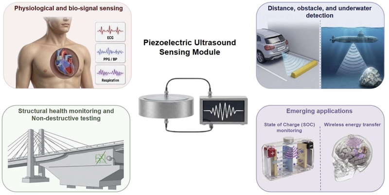
The categories of ultrasound-based piezoelectric sensors’ applications: bio-signal and physiological sensing, air/water distance and obstacle detection, structural health monitoring (SHM) and non-destructive testing (NDT), and emerging uses such as battery-state and wireless energy transfer.
2. KEY PIEZOELECTRIC PARAMETERS FOR ULTRASOUND SENSORS
At the core of an ultrasound-based piezoelectric sensor is the piezoelectric effect, so the performance of the sensing module is dictated by a compact set of material constants: the piezoelectric charge coefficient (d) and voltage coefficient (g), the electromechanical coupling factor (k), and the mechanical quality factor (Qm). The charge coefficient, defined as the change in electric displacement (D) per unit stress (T) at constant electric field (E), [32]
| (1) |
sets how much electrical charge an incident acoustic pressure liberates, so that in the linear constitutive description the electric displacement under an applied stress is written as [33]
| (12) |
where εT is the permittivity at constant stress. As the polarization generated at the material level is transferred through an actual circuit to operate in specific applications, g is an important parameter, defined at constant electric displacement, and can also be obtained by dividing d by permittivity [32].
| (3) |
The other constants describe conversion quality. k represents the square root of the fraction of input electrical (or mechanical) energy converted to output mechanical (or electrical) energy and thus determines the achievable sensitivity and bandwidth of the sensor [34]. Qm represents the inverse of mechanical loss at resonance, which is related to heat dissipation in practical device operation [35].
Various techniques have been developed to enhance these factors. The first family of strategies targets d itself, together with k, since k² = d²/(sEεT) rises with d so long as the elastic compliance and permittivity paid in return stay bounded [36]. The most general method is compositional tuning to a morphotropic phase boundary (MPB) or polymorphic phase boundary (PPB) where the coexistence of multiple phases provides a relatively large number of available polarization directions, leading to superior piezoelectric performance. This strategy is frequently coupled with other microstructural approaches such as texturing, secondary sintering, and dopant engineering to maximize piezoelectric performance across various systems [37-40]. Dopant engineering supplies a second strategy with a different approach. Applying donor doping by substituting higher valence causes metal vacancies and promotes movement of domain walls, which can lead to higher d [41]. Recently, rare-earth dopants (Sm3+, Eu3+, Sc3+, La3+, Er3+, etc.) act as inherent donors on A-sites, while additionally inducing local structural heterogeneity that fragments long-range polar order into nanoscale regions, and the resulting random fields lower the energy cost of polarization rotation [42]. Sm3+-doped PMN-PZT crystal ceramics near the MPB achieve exceptionally high performance (d33 = 910 pC/N, k33 = 0.81) [43]. In another approach, Eu3+ substitution onto Pb2+ sites in PIN-PMN-PT single crystals induces local lattice distortions and polar nano-regions (PNRs), boosting d33 from 2110 to 2870 pC/N alongside full-matrix anisotropy characterization [44]. Furthermore, Sm/Pr co-doped 0.72PMN-0.28PT ceramics push d33 up to 1310 pC/N, suggesting that strategically combining dual dopants can further enhance piezoelectric properties [45]. Templated grain growth (TGG) utilizes aligned templates to induce epitaxial growth of matrix grains during sintering, resulting in highly textured polycrystalline ceramics. This specific crystallographic alignment enables the bulk ceramic to mimic single-crystal domain behavior, offering a solution to achieve enhanced piezoelectric properties. Eu-doped PMN-PZT textured with 3 vol% BaTiO3 templates reaches d33 = 1310 pC/N and k33 = 0.87, against 325 pC/N and 0.70 for the random ceramic [46]. Nor is the approach confined to lead-based systems: a lead-free NKNS-BZ-BAZ ceramic textured with NaNbO3 templates to a Lotgering factor of 98.4% delivers d33 = 805 pC/N [47].
For sensors that combine transmit and receive functionality in a single element, the figure of merit (FoM) is commonly expressed as d × g, reflecting the combined contribution of the transmitting (d) and receiving (g) capabilities [48]. However, based on Eq. (3), d and g cannot be optimized independently since compositions engineered for a large d invariably carry a high permittivity, which suppresses g. To bypass this counterproductive effect, more sophisticated strategies aim to increase d that is not accompanied by a corresponding rise in permittivity [49]. In lead-free systems, textured KNNS-BNZ-BF combining a compositional design for structural flexibility with grain-oriented texturing yields a d33 ~ 807 pC/N and FoM ~ 64.7 × 10⁻¹² m2/N [50]. A similar strategy was demonstrated in the relaxor-PT single crystals by introducing large-radius Ho3+ ions at the B-site to increase ion mobility and oxygen-octahedral distortion. This suppresses the rise in εr typically accompanying doping-induced enhancement of d33, resulting in an FoM of 199 × 10-12 m2/N [51]. Table 1 summarizes the material properties of the various piezoelectric compositions mentioned above.

Piezoelectric material characteristics for ultrasound-based devices in various applications
In applications such as high-frequency pulse-echo and high-power transmit-receive modules, Qm becomes the key limiting factor because energy dissipated through internal mechanical loss is converted to heat, which depresses both transmitted pressure and echo signal-to-noise ratio (SNR) [52]. The standard route is the mirror image of the donor chemistry above: acceptor doping with low-valence cations such as Mn, Cu, Fe, whose charge-compensating oxygen vacancies bind to the acceptor into defect dipoles that establish an internal bias field and pin domain-wall motion. Mn-doped 0.15PYN-0.48PMN-0.37PT raises Qm to 1159 while holding d33 at 345 pC/N and Tc at 207℃ [53]. The same chemistry now extends to lead-free systems, where Cu-doped defect dipoles at an orthorhombic-tetragonal phase boundary in (K, Na)NbO3 increase Qm fourfold at the cost of only a one-fifth reduction in d33 [54]. Because pursuing hardening alone suppresses domain wall mobility via defect dipoles and ultimately limits d, recent studies have overcome this trade-off by combining multiple mechanisms acting on different length scales [55]. Superimposing texture onto defect engineering has proven to be the most effective strategy. For instance, in [001]-textured 9PMnN-91PZT, Mn lowers the sintering temperature sufficiently to preserve the BaTiO3 templates, combining d33 = 360 pC/N with Qm = 2507 at Tc = 261℃ [56]. Complementary doping achieves the same reconciliation chemically. Co-doping an A-site Eu donor and a B-site Mn acceptor in textured 0.24PIN-0.46PMN-0.30PT spatially separates the cation-oxygen vacancy recombination that would otherwise cancel the opposing effects. This dual strategy delivers d33 = 508 pC/N alongside kp = 0.64, Qm > 850 [57]. The material properties of the previously discussed piezoelectric compositions are outlined in Table 2. In practice, the relative importance of these parameters during sensor design varies depending on application constraints and operating environments. For instance, in biomedical applications that require the detection of weak signals, the parameter g, which is directly related to sensor sensitivity, is usually prioritized. Furthermore, in defect inspection applications where a single sensor consists of a transmitting and a receiving module, both parameters d and g are generally considered. Qm also becomes a critical parameter to evaluate, particularly in high-power devices prone to significant heat generation.
Piezoelectric material characteristics for high-power ultrasound-based devices.
3. APPLICATIONS
3.1 Physiological and bio-signal sensing
Biomedical reception is defined by a compromise between penetration and resolution. Its interrogation is typically performed in the megahertz (MHz) range. For example, deep organs such as the heart are probed at 2-5 MHz, and superficial vessels and skin structures require 10 MHz or higher [7,58,59]. Since the acoustic attenuation coefficient in tissue rises largely with the increase of incident acoustic frequency, the receiving module in a sensor must maintain high sensitivity across a wide frequency band as dictated by the target [60]. For this reason, two main factors dominate the design of the sensor. The first is the acoustic impedance matching. The acoustic impedance of the active element should match that of biological tissue (~1.5 MRayl) to minimize boundary reflection and maximize the acoustic coupling with the sensor [61]. To achieve this impedance matching, low-impedance materials such as PVDF or polymer-rich composites are widely adopted in wearable acoustic sensors [62]. Second, as discussed in Section 2, materials with a high g are preferred to maximize the generated voltage even under weak pressure waves. Based on these criteria, wearable sensors are designed for specific physiological targets. The target type and depth largely determine the choice of frequency, material, and array format.
1–3 composites are widely used in biomedical applications because incorporating a low-impedance polymer matrix lowers their effective acoustic impedance toward that of biological tissues, greatly enhancing acoustic coupling efficiency [63]. Furthermore, their inherent flexibility accommodates human body movements, thereby improving mechanical durability and extending the lifespan of the device. A prominent example of biomedical sensing that leverages these tailored material properties is hemodynamic monitoring by ultrasound wall tracking, which converts wall echo delay into a vessel diameter to yield blood pressure waveforms, as illustrated in Fig. 2(a) [64]. A 240 µm-thick conformal 1-3 composite array stretchable to 60% recovered central blood pressure waveforms from the carotid artery and jugular veins [8], and a flexible PZT-5A/PDMS 1-3 composite sensor with silver-nanowire electrodes captured ulnar-artery waveforms without occlusion or calibration [65]. Subsequent studies utilized advanced methodologies to further elevate wearability and measurement accuracy. For instance, a 4 × 5 array hot-press-transferred onto breathable fabric through “S”-shaped island-bridge electrodes achieved an axial resolution of 330 µm, while maintaining a measurement error of ± 2 mmHg compared to a commercial reference (Fig. 2(b)) [66]. The 45% ceramic filling factor of the PZT 5H 1-3 composite serves as a key tuning parameter, lowering the acoustic impedance to 15.3 MRayl for better tissue coupling while retaining a high thickness-mode coupling coefficient of kt = 0.62. Similarly, a 5 × 4 PMN-PT 1-3 composite array integrated via low-temperature Sn–Bi eutectic solder operated at 6.0 MHz with a 45° acceptance angle, tracking pressure within 4 mmHg of a commercial standard (Fig. 2(c)). The device leverages the high d33 (1282 pC/N) of PMN-PT to achieve signal accuracy without any matching or backing layers, overcoming the acoustic impedance mismatch (16.5 MRayl to tissue). Additionally, the sub-140°C soldering process was critical to strictly keeping the fabrication temperature below the PMN-PT Curie temperature, preventing any degradation of its piezoelectric properties [64].
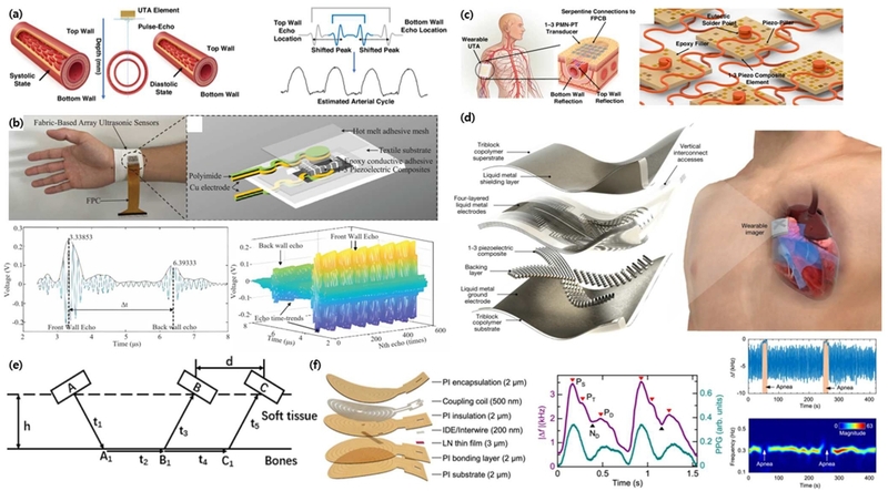
Representative ultrasound-based piezoelectric sensors for biomedical applications. (a) Schematic illustration of hemodynamic monitoring based on pulse-echo ultrasound signals reflected from arterial walls. Adapted from Ref. [64]. (b) A breathable, fabric-integrated ultrasound array utilizing "S"-shaped island-bridge electrodes for accurate continuous blood pressure measurement. Adapted from Ref. [66]. (c) Conceptual illustration of a wearable PMN-PT composite array for precise pressure tracking, paired with a detailed 3D rendering that highlights its serpentine connections and eutectic solder joints. Adapted from Ref. [64]. (d) Exploded structural view of a stretchable piezocomposite array incorporating liquid-metal electrodes, and an illustration of its application for dynamic cardiac imaging. Adapted from Ref. [69]. (e) Geometric schematic illustrating the transmission and reflection pathways of ultrasound-guided waves through soft tissue and bone for multi-site bone densitometry. Adapted from Ref. [70]. (f) Exploded diagram showing the functional layers of a wireless, suspended lithium niobate (LN) thin-film sensor, accompanied by in vivo measurement graphs tracking physiological signals and apnea events. Adapted from Ref. [71].
Biological imaging places rigorous requirements on coupling stability and channel count, as high-resolution reconstruction requires precise, phase-distortion-free beamforming [67,68]. To achieve this without compromising the high performance of conventional rigid transmitting modules, one strategy optimizes the acoustic interface using auxiliary coupling agents. For example, a bio-adhesive hydrogel–elastomer layer provided long-term acoustic matching, enabling a rigid probe to achieve 48 hours of continuous imaging across internal organs [58]. Additionally, integrating 1-3 piezocomposites with an acoustic matching layer improves both conformability and signal transmission. For instance, using Eco-flex as a matching layer bridges the impedance gap between the active element and human tissue, effectively minimizing wave reflection. A backing layer with a high attenuation coefficient effectively minimized the ringing effect, ensuring more accurate signal detection. By applying these integrated approaches, the sensor successfully achieved highly accurate imaging of the left ventricle during exercise (Fig. 2(d)) [69].
Beyond conventional vertical pulse-echo sensing, recent advancements have expanded into complex acoustic modes to address structural and physical constraints. For hard-tissue diagnostics, Fig. 2(e) demonstrates a rare-earth-doped PMN-PZT array (d33 = 525 pC/N) that utilizes axial-transmission guided waves for bone densitometry across the humerus, femur, and fibula. In this system, La/Sm co-doping raises k33 to 0.77, significantly outperforming undoped PMN-PT or PZT. This enhanced coupling efficiency sharpens the transmitted pulse and improves the timing precision, which is critical for accurate speed-of-sound measurements. The system employs three arrays tilted at 17°, 20°, and 23°, eliminating the need for strict normal-incidence geometry to accommodate the varying curvatures of different bone sites [70].
Furthermore, to overcome the weak-echo limitation inherent to deep or subtle physiological measurements, resonant operation offers a transformative pathway. Instead of listening for returning reflections, Fig. 2(f) depicts a wireless, battery-free 3-µm suspended lithium-niobate thin film sensor that tracks resonance-frequency shifts under mechanical deformation. This mechanism enables precise monitoring of soft arterial pulses and sleep apnea as a skin patch, while achieving intracranial pressure with 0.15 mmHg precision across a 240 mmHg dynamic range [71].
3.2 Distance, obstacle, and underwater detection
Distance measurement and obstacle detection are essential technologies across various application fields. They are fundamentally based on the ToF principle. In other words, physical position is calculated by receiving a signal emitted from a transmitter after it reflects off a target and precisely measuring the elapsed time [72]. To ensure sensor reliability in complex and diverse measurement environments, it is crucial to minimize signal scattering or attenuation caused by the medium and to clearly extract only the desired target signal. Compared to widely used optical or radio frequency (RF)-based sensors, ultrasound sensors are unaffected by the optical or electromagnetic properties of the surrounding medium. While laser sensors can barely detect transparent targets, ultrasound sensors are capable of stable sensing regardless of color or transparency [73]. For instance, a 3 × 3 PMUT array sensor located various transparent targets within 0.5 mm, as shown in Fig. 3(a) [74]. The cost is also quite low, as a sputtered PZT film delivered 0.764 Pa/V and an 87.4 Pa array output at 52 kHz [75]. Additionally, while detection resolution improves as frequency increases, signal attenuation within the medium also increases with frequency [76]. Considering this trade-off between resolution and detection range, utilizing piezoelectric sensors, which have relatively high electromechanical coupling characteristics, is the most effective approach among ultrasound-based signal detection devices.
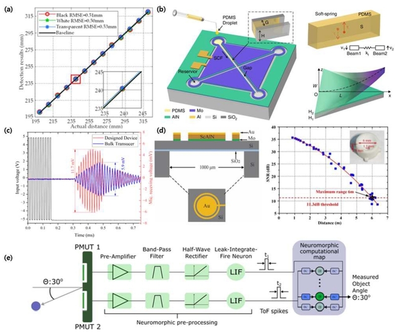
Ultrasound-based piezoelectric sensors for airborne distance measurement and obstacle detection. (a) A linear regression plot illustrating the highly precise spatial localization performance of a PMUT array, even for transparent targets. Adapted from Ref. [74]. (b) Isometric schematics, cross-sectional views showing device layers, and corresponding mechanical models for a cantilever-based PMUT architecture utilizing soft polymer interconnects. Adapted from Ref. [80]. (c) Transient response graphs showing input and receiving voltage plots, demonstrating significantly enhanced sensitivity for a ScAlN PMUT array compared to conventional bulk transducers. Adapted from Ref. [88]. (d) Schematic cross-sections (top-left), top-down views (bottom-left), and a signal-to-noise ratio (SNR) plot (right) with a device inset photo, outlining a structurally optimized single-element dual-ring ScAlN device for long-range measurement. Adapted from Ref. [89]. (e) A complete system block diagram illustrating the neuromorphic signal processing flow, where paired PMUT echoes are pre-processed and converted into spike-based ToF signals. Adapted from Ref. [90].
In the design of air-coupled sensors, the most critical challenge is overcoming the enormous acoustic impedance mismatch, spanning approximately four orders of magnitude, between the sensor and air. Without proper impedance-matching strategies, most of the incident acoustic energy is reflected at the sensor–air interface, severely degrading both the transmitted acoustic pressure and the sensor sensitivity. To address this issue, various strategies have been developed. The most common approach is to insert a quarter-wavelength matching layer with very low acoustic impedance, such as porous polymers, hollow-microsphere composites, or silica aerogels, between the sensor and air [77,78]. For instance, a hollow polymer microsphere/epoxy matching layer improved the sensor sensitivity by 20.9 dB [79]. A cantilever PMUT with soft polymer interconnects that synchronize neighboring cells and act as a reflection-reduction agents covered 0.27–3.8 m over a 170° field (Fig. 3(b)) [80]. However, since the ideal matching impedance for air is on the order of 0.02 MRayl, a single layer is often insufficient, and multilayer or graded-impedance structures have been proposed to provide a more gradual impedance transition [81]. A 1–3 piezocomposite air-coupled sensor achieved a two-way insertion loss of −56.86 dB by utilizing a double-layer matching structure made of hollow glass microsphere-filled epoxy and a micro-membrane filter [82]. Building on this concept, traditional discrete layers can be replaced by a continuous impedance gradient. For instance, a cone-structured acoustic metamaterial using silica-epoxy unit cells was developed [83]. By gradually reducing the silica content away from the piezoelectric element, they lowered the acoustic impedance from 11.4 to 3.0 MRayl and enabled an exceptionally broad −6 dB bandwidth of over 100%. This highlights a key advantage of acoustic metamaterials, which control impedance through their structural geometry rather than just material composition. Consequently, even exceptionally thin metasurfaces with resonators or air cavities can maximize wave transmission. An ultrathin metasurface (λ/100) designed for the water-air interface demonstrated a 100-fold increase in acoustic transmission, from 0.1% to approximately 30% [84]. In addition to addressing the impedance mismatch at the interface, macroscopic structural designs such as horn-type acoustic concentrators or focused acoustic lenses offer complementary approaches to further improve efficiency [85,86]. By physically focusing acoustic waves to amplify local pressure, these structures directly compensate for another major challenge in air-coupled ultrasound, which is extreme signal attenuation. Indeed, in terms of attenuation, while water (0.0022 dB/cm/MHz) and soft tissue (0.75 dB/cm/MHz) exhibit low attenuation coefficients, air demonstrates an extremely high signal attenuation coefficient of 7.50 dB/cm/MHz [87]. Therefore, to effectively implement an air-coupled ultrasound sensor system, the receiving sensitivity of the sensor must be maximized by selecting a piezoelectric material with a high piezoelectric coefficient, enabling it to output a high voltage even from severely attenuated, minute echo signals. Scandium-doped AlN (ScAlN) is highly advantageous here, as its low permittivity yields a favorable g, boosting receive voltage. Demonstrating this, a ScAlN PMUT array doubled the acoustic pressure and receiving voltage (11.7 vs 5.9 mV at 10 cm) of bulk transducers, achieving a range beyond 6.8 m (Fig. 3(c)) [88]. Similarly, a single-element dual-ring ScAlN device (kt2 = 8.92%) achieved a 6 m range (Fig. 3(d)) [89]. Recently, spatial detection advanced through neuromorphic read-outs (Fig. 3(e)), where a pair of PMUTs converts acoustic echoes into spike-based ToF signals. Resistive-memory coincidence detectors then map these arrival time differences into precise target angles [90].
Ultrasound-based distance and obstacle sensors offer significant advantages, particularly in underwater environments, as conventional electromagnetic and optical detection technologies are inherently limited by the high absorption and scattering properties of water [91]. The first man-made echo-ranging device, Fessenden’s 540 Hz electrodynamically driven oscillator, demonstrated depth sounding and returned an echo from an iceberg at a distance of 3,200 m in March 1914 [92]. The application of piezoelectricity in this field began during World War I, when Langevin pioneered the sandwich resonator, which is an assembly of quartz plates bonded between steel masses to detect submerged submarines [93]. Following the commercial introduction of the fathometer in the mid-1920s, its application expanded rapidly within a decade. Shortly after acoustic echoes from cod were identified in sound traces, fish-finding sonars entered the market in 1948 [94]. Building upon this foundational work, numerous subsequent technical studies to improve sensitivity have been conducted (Fig. 4(a)) [95]. For example, a matching-layer PMN–PT rod sensor improved free-field receiving voltage sensitivity by over 6 dB relative to its PZT-4 counterpart [96]. As illustrated in Fig. 4(b), ordered polar micro-holes machined into [001]-oriented PIN-PMN-PT pillars introduce a dual-piezo-charge mechanism that raises the sensitivity compared to previous approaches [97].
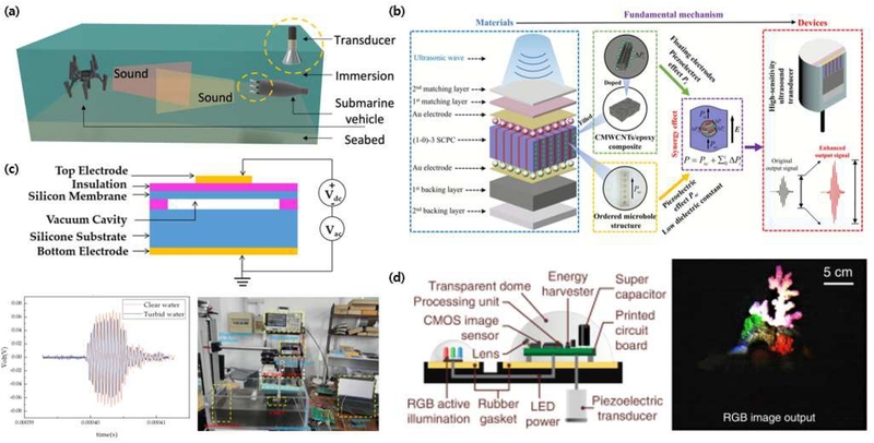
Ultrasound-based piezoelectric sensors for underwater distance measurement and obstacle detection. (a) A 3D conceptual schematic representing foundational underwater acoustic sensing, illustrating ongoing technological advancements in high-sensitivity sonar systems for submarine vehicle obstacle detection. Adapted from Ref. [95]. (b) Detailed exploded structural schematics and fundamental mechanism diagrams of an advanced transmitting module of ultrasound sensors, where ordered polar micro-holes machined into PIN-PMN-PT pillars introduce a dual-piezo-charge mechanism to significantly raise sensitivity. Adapted from Ref. [97]. (c) A transient waveform plot comparing signals in clear and turbid water (top-left) alongside a cross-sectional device schematic (bottom) of a PDMS-encapsulated CMUT. This architecture provides robust and stable ranging performance for robotic fish in visually opaque environments where optical methods completely fail. Adapted from Ref. [98]. (d) An underwater RGB photograph (left) and a comprehensive system cross-section diagram (right) of a fully submerged, battery-free camera. The integrated piezoelectric transducer system enables ultra-low-power localization, backscatter communication, and imaging. Adapted from Ref. [101].
Recent research has focused on the performance requirements of underwater sensors are required to perform. Miniaturized MEMS sonars are now being mounted on platforms too small for conventional projectors. While piezoelectric sensors remain the primary focus, non-piezoelectric capacitive micromachined ultrasound transducers (CMUT) provide a crucial complementary benchmark for these advanced underwater sensing platforms. For a robotic fish to track obstacles within a long-distance range (10 to 42 cm), a PDMS-encapsulated CMUT was mounted and enabled its ranging error to remain unchanged even when sand and ink were added to simulate turbid water: a condition where optical ranging completely fails (Fig. 4(c)) [98]. Pushing toward resolution rather than range, a CMUT with a 100% fractional bandwidth resolves 4 mm targets and reconstructs the topography of submerged 3D structures up to 60 cm away, even operating at a relatively high frequency of 1.5 MHz [99]. The most remarkable achievement is the dramatic reduction in power consumption. These piezoelectric nodes harvest energy from incoming sound waves and communicate simply by changing their own impedance, which drastically lowers the underwater power consumption. In real-world river tests, metamaterial-enhanced nodes maintained their speed over 62 m and successfully connected more than 10 devices. Compared to previous state-of-the-art technologies, this represents a 5-fold increase in data speed and a 6-fold increase in transmission range [100]. Built on the same principles, a fully submerged battery-free camera localizes tags with sub-10 cm accuracy at 3.5 m and decodes a 1 kbps uplink from beyond 40 m, while shooting images while consuming an average power of 276.31 µW, which is 5 times lower than previous underwater wireless imagers, with the backscatter modulation itself drawing just 24 nW (Fig. 4(d)) [101]. Taken together, the air and underwater cases show how FoM reorders the entire hierarchy of material and architecture choices.
3.3 Structural health monitoring and non-destructive testing
In structural health monitoring applications, the sensor is bonded to, or embedded in, the component. It detects guided elastic waves, which are mechanical stress waves that propagate along the physical boundaries of a structure to enable long-range damage detection [102]. The mode, velocity, and arrival time reveal the presence and location of hidden damage. Directly integrated into the structure, the active sensors transfer mechanical strain without intermediate coupling media. To successfully capture weak scattered signals from distant damage, their design must ensure stable bonding, precise mode selectivity, and high piezoelectric sensitivity. Several papers detail how specific sensor geometries, wave-mode strategies, and signal-processing chains have made this approach the mainstay of structural diagnostics [103-106]. The concept of piezoelectric wafer active sensors (PWAS) using a bonded PZT wafer to excite and detect tuned Lamb waves was originally established for aircraft-grade aluminum plates. This work demonstrated S0 mode using a PWAS and pulse-echo crack detection over distances up to 2,500 mm using a phased sensor array [107]. Today, high-coupling PMN-PT 1-3 composites (d33 = 1208 pC/N, kt = 0.857) provide the broadband, high-sensitivity sensors needed for ToF diffraction defect detection (Fig. 5(a)) [34].
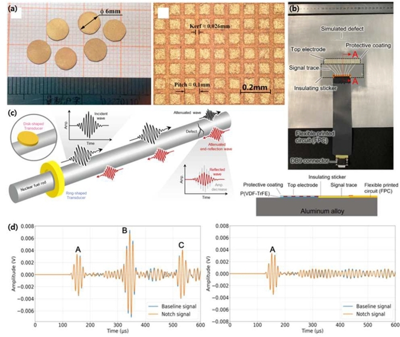
Ultrasound-based piezoelectric sensors for structural health monitoring and non-destructive testing. (a) Macroscopic optical photographs (left) and a microscopic surface image (right) of high-coupling PMN-PT 1-3 composites, which serve as broadband, high-sensitivity sensors for ToF diffraction defect detection. Adapted from Ref. [34]. (b) A top-down photograph (top) and a corresponding cross-sectional schematic (bottom) of a direct-write sensing platform applied to an aluminum plate, configured to resolve small structural defects against acoustic edge echoes. Adapted from Ref. [109]. (c) A 3D conceptual schematic accompanied by transient wave propagation graphs, illustrating a direct-ink-written, Sm-doped PMN-PT ring-type sensor conforming tightly to curved, narrow-access pipes for continuous defect monitoring. Adapted from Ref. [110]. (d) A transient voltage response graph with an inset photograph (left) alongside a 3D structural schematic (right) of a thin, flexible piezoelectric array. This highly conformable architecture enables phased-array focusing to pinpoint defects on curved or confined aerospace components. Adapted from Ref. [112].
A major challenge with rigid sensors is their poor adhesion to curved or irregular surfaces, which often leads to detachment under operational stress. To address this, recent research has heavily focused on developing conformal, directly integrated receiving modules for sensors. For instance, direct-write P(VDF–TrFE) sensors deposited straight onto pipe walls detected simulated defects as shallow as 0.25 mm and resolved wall-thickness changes with under 3% error via pulse-echo timing [108]. Building on this same direct-write sensing platform, a thinner coating on an aluminum plate similarly resolved a 1 mm deep, 2 mm wide defect against the S₂ mode edge echo (Fig. 5(b)) [109]. The limited access and fixed geometry of pipeline monitoring have driven the development of dedicated architectures. Additive manufacturing directly addresses these physical constraints. For instance, a direct-ink-written, Sm-doped PMN-PT ring-type sensor can conform tightly to curved, narrow-access pipes (Fig. 5(c)). Because it is excited uniformly around its circumference, it generates purely axisymmetric guided waves, which eliminate the circumferential interference that degrades conventional disk-type probes in pulse-echo detection, achieving a signal-to-noise ratio of 41.1 dB, nearly 7-fold above the 6 dB reliability threshold [110]. Additionally, for real-time processing, FPGA-based smart nodes leverage the torsional mode to localize pipe defects on-board with errors as low as 8 mm, just 2% of the 400 mm sensing span [111].
Furthermore, civil and aerospace structures present their own unique constraints, which require highly specialized sensor designs. For large-scale framework monitoring, guided-wave sensor arrays combined with multivariate outlier analysis can reliably inspect fatigue-prone truss welds, achieving up to 100% damage detection efficiency with the best-performing sensing path, which is further detailed in Fig. 5(d) [112]. As spatial constraints become more severe on curved or confined aerospace components, maintaining conformability becomes paramount: thin, flexible piezoelectric arrays enable phased-array focusing, such as concentrating A₀ mode waves over a 100 mm path to pinpoint 10 mm defects [113]. Likewise, when high-resolution imaging on planar components is required without wave dispersion, SH₀ wave phased arrays offer an ideal alternative, capturing through-thickness flaws as small as 2 mm [114]. Across all these applications, the sensor requirements differ fundamentally from traditional imaging: they demand moderate frequencies, high sensitivity to faint scattered waves, and, above all, a coupling interface that remains stable over the structure's lifetime.
3.4 Emerging applications
While the applications examined so far, like biosensing, obstacle and distance detection, and non-destructive testing, have primarily focused on identifying macroscopic shapes or defect locations by utilizing the reflective properties of ultrasound, new fields of application have recently emerged that maximize the inherent advantages of ultrasound as a mechanical wave [13]. In sealed environments impenetrable to light or electromagnetic waves, ultrasound serves as an effective method for traversing physical barriers. By leveraging the characteristics of mechanical waves that respond to minute changes in the medium, ultrasound can function as a sensor to monitor the physical and chemical states of a closed system.
With the recent surge in the use of high-capacity lithium-ion batteries, the early detection of thermal runaway, which can directly lead to fires, and the accurate monitoring of State of Charge (SOC) and State of Health (SOH) have become critical [115,116]. However, conventional methods relying solely on external voltage and current measurements are limited in their ability to detect localized structural changes or side reactions within the internal electrodes. In contrast, ultrasound waves can penetrate the battery casing and possess unique characteristics whereby the signal is modulated by internal chemical and physical states as the mechanical waves propagate through the medium [117]. For instance, the lithiation process of an electrode alters the internal speed of sound, while gas generation resulting from battery degradation induces significant acoustic scattering. Consequently, the internal state can be estimated by measuring the ToF and attenuation of ultrasound waves [118]. Piezoelectric materials play a crucial role in implementing such diagnostic systems. Due to the high electromechanical coupling characteristics of piezoelectric elements, operation is possible with a single element, and miniaturization or thinning is easily achieved. This enables real-time monitoring through direct contact with the surface of a commercial battery cell, without requiring any structural modifications. A single built-in piezoelectric sensor can estimate the SOC with a root-mean-square (RMS) error below 2.36% using direct guided waves [119]. More advanced phased-array systems can map the SOC to within 4% while visualizing gas generation inside a 40 Ah cell [120]. Similarly, reflection-based schemes can effectively estimate the battery’s charge and temperature, as depicted in Fig. 6(a) [121]. In addition, real-time imaging enables the non-destructive visualization of lithium plating as it forms (Fig. 6(b)) [122]. A recent passive ultrasound probe embedded in a cell provides early warning of internal thermal-runaway hazards, explicitly utilizing a PZT element (d33 = 350 pC/N, kt = 0.60) operating at 2.5 MHz [123]. In all these cases, the appeal of piezoelectric sensors is clear: they are inexpensive, compact, and easily attached to commercial cells without altering them.
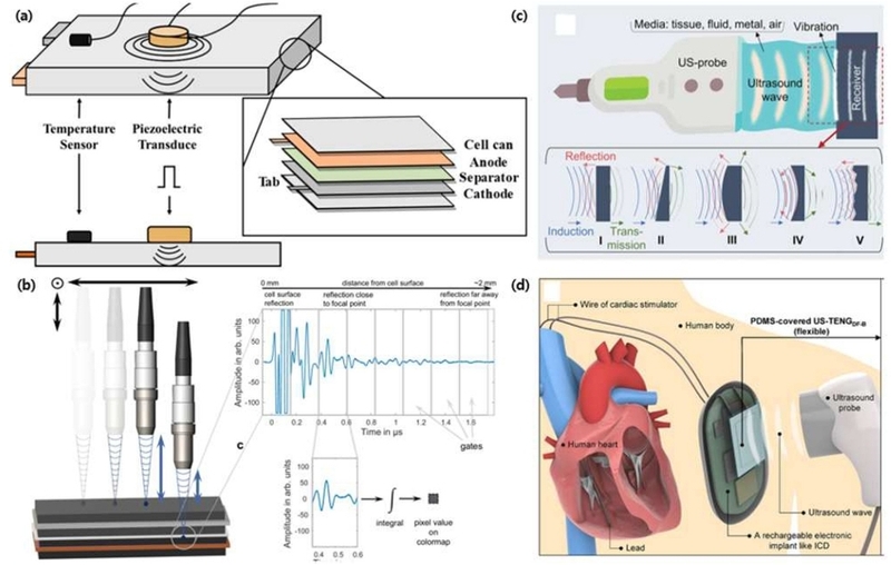
Emerging applications of ultrasound-based piezoelectric technologies. (a) A 3D structural schematic detailing a reflection-based ultrasound scheme designed to effectively estimate a battery's SOC and internal temperature across its constituent layers. Adapted from Ref. [121]. (b) A conceptual scanning diagram paired with transient acoustic reflection waveforms, demonstrating the capability for real-time, non-destructive visualization of lithium plating as it forms. Adapted from Ref. [122]. (c) A system schematic alongside wave propagation models illustrating ultrasound wireless energy transfer technology, highlighting acoustic transmission through various media. Adapted from Ref. [125]. (d) An anatomical application schematic of a flexible receiver on a human heart. Adapted from Ref. [127].
In addition to such state-sensing applications, the mechanical wave characteristics of ultrasound enable a new paradigm moving beyond mere information reception to the transfer of energy. Unlike conventional electromagnetic wave-based methods, ultrasound allows for the safe and efficient transmission of high-density mechanical energy even through various enclosed media without the risks of electromagnetic interference or heat generation [124]. Consequently, ultrasound wireless energy transfer technology based on these principles has recently been gaining renewed attention. As shown in Fig. 6(c), the structure and design of the receiver are important because diverse surface structures of the receiver change the direction and shape of the transmitted ultrasound waves [125]. For instance, a receiver utilizing a relaxor single crystal successfully transferred approximately 8 mW of power across 6 cm of water. Furthermore, it demonstrated the ability to deliver power through solid barriers like metal and wood [126]. A subsequent body-conformal design supplied reliable power to a rechargeable battery at a 35 mm distance. Notably, it maintained its output under repeated bending, proving its suitability for curved implants such as artificial hearts (Fig. 6(d)) [127]. With these demonstrations validating the immense potential of ultrasound-based energy transfer, piezoelectric materials have emerged as a suitable platform for this technology. Some groups have achieved significant power generation in vivo using purely piezoelectric receivers. An implantable harvester based on a Sm-doped PMN-PT single crystal produced 280 µW in rat brain tissue, which was sufficient to trigger a measurable analgesic response [128]. Additionally, recent millimeter-scale implants have successfully integrated both powering and sensing into a single ultrasound link. One such device continuously monitored deep-tissue oxygen levels up to a 10 cm depth by receiving power and transmitting data simultaneously via backscatter modulation [129].
What unites these emerging applications is a fundamental shift in the sensor's role. Rather than focusing on the high image quality required in medical and industrial ultrasound, these systems prioritize the ability to detect hidden state variables such as charge, gas, flow, or motion. No competing sensor can access these variables as directly or as cost-effectively.
4. CONCLUSION
Despite the broad applications surveyed in Section 3, surpassing current performance limits requires looking beyond traditional material optimization. While Section 2 outlined how intrinsic constants (d, g, k, and Qm) can be tuned through compositional engineering, modern wearable and implantable formats demand far more complex operational capabilities such as mechanical flexibility, biocompatibility, and lead-free compliance that single homogeneous ceramics cannot deliver alone. Since improving intrinsic material properties alone is no longer sufficient to meet modern demands, overcoming current performance limits requires innovative structural and system-level designs. Three complementary strategies are proposed.
The first strategy employs metamaterials to manipulate the incident waves. Metamaterials are engineered structures with properties not found in nature, enabling the artificial manipulation of waves. By introducing metamaterials specifically tailored for ultrasound, the efficiency of the sensor can be maximized. For instance, anisotropic metamaterial architectures can amplify pressure at the sensor interface by over an order of magnitude and enhance the SNR by more than 20 dB [28]. The approach extends further to energy-focusing metamaterial geometries tailored for energy harvesting and self-powered sensing [130]. Because these enhancements are purely geometric, they directly multiply whatever baseline sensitivity the material provides, establishing structural amplification as a powerful route for ultrasound-based sensors.
The second strategy focuses on redesigning active materials for direct use on or inside the body, moving beyond simple flexibility toward true tissue integration. Because of this direct contact with the human body, the evaluation criteria must expand. Beyond traditional sensitivity, essential requirements now include cytocompatibility, mechanical softness that matches human tissue, and ideally, controlled bio-resorption. Recent advances in biocompatible piezoelectric materials demonstrate that wirelessly activated ultrasound can deliver localized, minimally invasive electrical cues, establishing a viable bridge from laboratory concepts to practical clinical applications [131,132]. A representative milestone is an implantable, flexible lead-free device capable of generating focused acoustic pressure for target deep-brain stimulation while maintaining safety [133]. These multifunctional systems blur the boundary of a sensor, stimulator, and implant. Consequently, next-generation devices will be evaluated not only on their individual performance but also on their functional integration with living tissue.
Driven by environmental and biomedical regulations, the third strategy focuses on developing biocompatible and eco-friendly piezoelectric materials. Recent advances in KNN-based materials and processing have significantly enhanced their coupling and sensitivity, fulfilling the performance requirements for modern ultrasound-based devices. A KNN-based wearable imaging array achieved enhanced d33 from a single flexible probe [134]. Additionally, a KNN-composite with high g33 was used for receiving and energy harvesting [135]. Although KNN still exhibits a slight performance gap in parameters such as d and k compared to Pb-based ceramics like PZT [136,137], this difference is being overcome through advanced processing techniques such as single crystallization and templated grain growth, making it highly promising for future practical applications.
The performance of an ultrasound-based piezoelectric sensor is governed by a compact and well-understood set of FoM: the piezoelectric charge and voltage constants, the electromechanical coupling factors, the mechanical quality factor, the permittivity, and, above all, the acoustic impedance whose relative weight is decided by the medium into which the device must listen. These same parameters, traced through this review, explain why low-impedance polymers and dense arrays dominate wearable health monitors, why flexural micromachined membranes prevail in air-coupled ranging while bulk composites prevail underwater, why stably bonded wafer sensors define structural monitoring, and why inexpensive attached sensors are opening the sealed interiors of batteries and pipes to inspection. The remaining frontier is to surpass the material limits of the active element without sacrificing wearability or safety. The convergence of metamaterial amplification, biocompatible material design, and increasingly capable lead-free ceramics indicates that the next generation of ultrasound-based piezoelectric sensors will be simultaneously more sensitive, more benign, and more deeply integrated into the systems they are built to observe.
Acknowledgments
The authors acknowledge this research was supported by the National Research Foundation of Korea (NRF) funded by Ministry of Science and ICT (MSIT) (RS-2025-25441823 and RS-2026-25497859) and partially supported by Korea University.
REFERENCES
-
T.-G. La, L.H. Le, Flexible and Wearable Ultrasound Device for Medical Applications: A Review on Materials, Structural Designs, and Current Challenges, Adv. Mater. Technol. 7 (2022) 2100798.
[https://doi.org/10.1002/admt.202100798]

-
C. Kollmann, Diagnostic Ultrasound Imaging: Inside Out (Second Edition), Ultrasound Med. Biol. 41 (2015) 622.
[https://doi.org/10.1016/j.ultrasmedbio.2014.09.026]
-
R. Horstmeyer, H. Ruan, C. Yang, Guidestar-Assisted Wavefront-Shaping Methods for Focusing Light into Biological Tissue, Nat. Photonics 9 (2015) 563–571.
[https://doi.org/10.1038/nphoton.2015.140]
-
V. Ntziachristos, Going deeper than microscopy: the optical imaging frontier in biology, Nat. Methods 7 (2010) 603–614.
[https://doi.org/10.1038/nmeth.1483]
-
K.K. Shung, Diagnostic Ultrasound: Imaging and Blood Flow Measurements, second ed., CRC Press, Boca Raton, 2015.
[https://doi.org/10.1201/b18323]
-
F.A. Duck, Medical and Non-Medical Protection Standards for Ultrasound and Infrasound, Prog. Biophys. Mol. Biol. 93 (2007) 176–191.
[https://doi.org/10.1016/j.pbiomolbio.2006.07.008]
-
M. Lin, Z. Zhang, X. Gao, Y. Bian, R.S. Wu, G. Park, et al., A Fully Integrated Wearable Ultrasound System to Monitor Deep Tissues in Moving Subjects, Nat. Biotechnol. 42 (2024) 448–457.
[https://doi.org/10.1038/s41587-023-01800-0]
-
C. Wang, X. Li, H. Hu, L. Zhang, Z. Huang, M. Lin, et al., Monitoring of the Central Blood Pressure Waveform via a Conformal Ultrasonic Device, Nat. Biomed. Eng. 2 (2018) 687–695.
[https://doi.org/10.1038/s41551-018-0287-x]
-
R.J. Przybyla, S.E. Shelton, A. Guedes, I.I. Izyumin, M.H. Kline, D.A. Horsley, et al., In-Air Rangefinding With an AlN Piezoelectric Micromachined Ultrasound Transducer, IEEE Sens. J. 11 (2011) 2690–2697.
[https://doi.org/10.1109/JSEN.2011.2157490]
-
A. Raghavan, C.E.S. Cesnik, Review of Guided-Wave Structural Health Monitoring, Shock Vib. Dig. 39 (2007) 91–114.
[https://doi.org/10.1177/0583102406075428]
-
S. Zhang, F. Li, X. Jiang, J. Kim, J. Luo, X. Geng, Advantages and Challenges of Relaxor-PbTiO₃ Ferroelectric Crystals for Electroacoustic Transducers – A Review, Prog. Mater. Sci. 68 (2015) 1–66.
[https://doi.org/10.1016/j.pmatsci.2014.10.002]
-
K.-Y. Park, Y.-B. Han, H.-J. Yoon, Triboelectrification-Based Ultrasound Detection, J. Sens. Sci. Technol. 34 (2025) 488–497.
[https://doi.org/10.46670/JSST.2025.34.5.488]
-
P. Manchi, H.-J. Yoon, Electrospun Nanofibers Based Flexible Triboelectric Nanogenerators for Mechanical Energy Harvesting and Smart Sensing Applications: A Review, Trans. Electr. Electron. Mater. 27 (2026) 90–99.
[https://doi.org/10.1007/s42341-025-00689-7]
-
Z. Ghemari, A. Bendaikha, S. Belkhiri, S. Saad, Fabrication of Flexible Resistive Pressure Sensors Using Graphene/Polydimethylsiloxane Composites, Trans. Electr. Electron. Mater. 26 (2025) 323–344.
[https://doi.org/10.1007/s42341-025-00601-3]
-
Y. Qiu, J.V. Gigliotti, M. Wallace, F. Griggio, C.E.M. Demore, S. Cochran, et al., Piezoelectric Micromachined Ultrasound Transducer (PMUT) Arrays for Integrated Sensing, Actuation and Imaging, Sensors 15 (2015) 8020–8041.
[https://doi.org/10.3390/s150408020]
-
Z. Ghemari, S. Belkhiri, Exploring Novel Piezoelectric Materials with Enhanced Thermal Stability for High-Performance Sensors, Trans. Electr. Electron. Mater. 26 (2025) 289–304.
[https://doi.org/10.1007/s42341-025-00600-4]
-
Q. Zhou, S. Lau, D. Wu, K. Kirk Shung, Piezoelectric Films for High Frequency Ultrasonic Transducers in Biomedical Applications, Prog. Mater. Sci. 56 (2011) 139–174.
[https://doi.org/10.1016/j.pmatsci.2010.09.001]
-
M. Tanveer, M.U. Elahi, J. Jung, M.M. Azad, S. Khalid, H.S. Kim, Recent Advancements in Guided Ultrasonic Waves for Structural Health Monitoring of Composite Structures, Appl. Sci. 14 (2024) 11091.
[https://doi.org/10.3390/app142311091]
-
H.-L. Yu, N.-R. Ko, W.-J. Choi, T.T. Zate, W. Jo, A Brief Review of Some Challenging Issues in Textured Piezoceramics via Templated Grain Growth Method, J. Sens. Sci. Technol. 32 (2023) 10–15.
[https://doi.org/10.46670/JSST.2023.32.1.10]
-
D.-G. Lee, S.-B. Kim, Y. Yan, S. Hur, H.-C. Song, Brief Review of Lead-Free Piezoelectric Ceramics for High-Power Applications, J. Sens. Sci. Technol. 34 (2025) 116–123.
[https://doi.org/10.46670/JSST.2025.34.2.116]
-
E.S. Lim, S.H. Lee, J. Choi, Y.J. Lim, H.U. Chung, Advances in Skin-interfaced Wireless Physiological Monitoring Systems, J. Sens. Sci. Technol. 34 (2025) 458–472.
[https://doi.org/10.46670/JSST.2025.34.5.458]
-
Y. Lee, S. Biswas, M. Koo, H. Kim, Recent Development in Biocompatible Biosensors, J. Sens. Sci. Technol. 32 (2023) 403–411.
[https://doi.org/10.46670/JSST.2023.32.6.403]
-
V. Giurgiutiu, Chapter 8 - Coupling of PWAS Transducers to the Monitored Structure, In: Structural Health Monitoring with Piezoelectric Wafer Active Sensors, second ed., Academic Press, Oxford, 2014, pp. 395–443.
[https://doi.org/10.1016/B978-0-12-418691-0.00008-3]
-
M. Mitra, S. Gopalakrishnan, Guided Wave Based Structural Health Monitoring: A Review, Smart Mater. Struct. 25 (2016) 053001.
[https://doi.org/10.1088/0964-1726/25/5/053001]
-
G. Davies, K.W. Knehr, B.V. Tassell, T. Hodson, S. Biswas, A.G. Hsieh, et al., State of Charge and State of Health Estimation Using Electrochemical Acoustic Time of Flight Analysis, J. Electrochem. Soc. 164 (2017) A2746–A2755.
[https://doi.org/10.1149/2.1411712jes]
-
Y. Gao, M. Chen, Z. Wu, L. Yao, Z. Tong, S. Zhang, et al., A Miniaturized Transit-Time Ultrasonic Flowmeter Based on ScAlN Piezoelectric Micromachined Ultrasonic Transducers for Small-Diameter Applications, Microsyst. Nanoeng. 9 (2023) 49.
[https://doi.org/10.1038/s41378-023-00518-y]
-
Z. Li, D.-Q. Yang, S.-L. Liu, S.-Y. Yu, M.-H. Lu, J. Zhu, et al., Broadband Gradient Impedance Matching Using an Acoustic Metamaterial for Ultrasonic Transducers, Sci. Rep. 7 (2017) 42863.
[https://doi.org/10.1038/srep42863]
-
Y. Chen, H. Liu, M. Reilly, H. Bae, M. Yu, Enhanced Acoustic Sensing through Wave Compression and Pressure Amplification in Anisotropic Metamaterials, Nat. Commun. 5 (2014) 5247.
[https://doi.org/10.1038/ncomms6247]
-
L. Jiang, Y. Yang, R. Chen, G. Lu, R. Li, D. Li, et al., Flexible Piezoelectric Ultrasonic Energy Harvester Array for Bio-Implantable Wireless Generator, Nano Energy 56 (2019) 216–224.
[https://doi.org/10.1016/j.nanoen.2018.11.052]
-
A.K. Das, P. Mishra, S.S. Biswal, A. Tripathy, S. Bhuyan, Processing and Development of Lead Free Bi(Ni₂/₃Nb₁/₃)O₃ Ferroelectric Material for Thermal Sensing Electronic Device, Trans. Electr. Electron. Mater. 27 (2026) 156–164.
[https://doi.org/10.1007/s42341-025-00646-4]
-
Y.-R. Choi, C.-I. Cheon, K.-W. Chae, Piezoelectric Strain Characteristics of BiFeO₃-BaTiO₃ Ceramics around the MPB Composition, J. Sens. Sci. Technol. 35 (2026) 216–224.
[https://doi.org/10.46670/JSST.2026.35.3.216]
-
D. Damjanovic, Ferroelectric, Dielectric and Piezoelectric Properties of Ferroelectric Thin Films and Ceramics, Rep. Prog. Phys. 61 (1998) 1267–1324.
[https://doi.org/10.1088/0034-4885/61/9/002]
-
S. Priya, H.-C. Song, Y. Zhou, R. Varghese, A. Chopra, S.-G. Kim, et al., A Review on Piezoelectric Energy Harvesting: Materials, Methods, and Circuits, Energy Harvest. Syst. 4 (2017) 3–39.
[https://doi.org/10.1515/ehs-2016-0028]
-
D. Liu, Q. Yue, J. Deng, D. Lin, X. Li, W. Di, et al., Broadband and High Sensitive Time-of-Flight Diffraction Ultrasonic Transducers Based on PMNT/Epoxy 1–3 Piezoelectric Composite, Sensors 15 (2015) 6807–6817.
[https://doi.org/10.3390/s150306807]
-
Z. Chen, L. Song, W. Cao, Characterization of High-Power Mechanical Quality Factor of Piezoelectric Ceramic Discs under Self-Heating Condition, J. Mater. Res. Technol. 23 (2023) 5040–5049.
[https://doi.org/10.1016/j.jmrt.2023.02.149]
-
X. Wu, D. Vanderbilt, D.R. Hamann, Systematic Treatment of Displacements, Strains, and Electric Fields in Density-Functional Perturbation Theory, Phys. Rev. B 72 (2005) 035105.
[https://doi.org/10.1103/PhysRevB.72.035105]
-
M. Wu, R. Yao, C. Jin, Y. Xu, J. Xu, L. He, et al., Significantly Enhanced Piezoelectric Properties of BaTiO₃-Based Ceramics with Unchanged Curie Temperature via Local Chemical Inhomogeneity, Chem. Eng. J. 518 (2025) 164844.
[https://doi.org/10.1016/j.cej.2025.164844]
-
F. Wang, T. Zhang, M. Guo, M. Zhang, Room Temperature Constructing Rhombohedral-Tetragonal Phase Boundary in Novel (Bi, Na)(Zr, Ti)O₃ Modified (K, Na)(Nb, Sb)O₃ Ceramics: Phase Structure, Defect and Piezoelectric Performance, Ceram. Int. 48 (2022) 19954–19962.
[https://doi.org/10.1016/j.ceramint.2022.03.270]
-
H. Song, Y.M. Jin, H. Kim, J. Shim, Y. Kim, C.-L. Lee, et al., Artificial Morphotropic Phase Boundary Associated with Nano-Clustering in Lead Zirconate – Lead Titanate Composite Films, Adv. Funct. Mater. 36 (2026) e25400.
[https://doi.org/10.1002/adfm.202525400]
-
G.-S. Lee, B.-J. Min, B.-H. Choi, H.-J. Choi, B. Kim, S. Nahm, Effect of Fe₂O₃ Additive on Structural and Piezoelectric Properties of (Na,K)(Nb,Sb)O₃–CaZrO₃–(Bi,Ag)ZrO₃ Lead-Free Piezoceramics, J. Mater. Chem. C 13 (2025) 22567–22579.
[https://doi.org/10.1039/D5TC02761F]
-
T.W. Kim, G. Lee, M. Ichimura, J.-H. Koh, Enhanced Soft Piezoelectric Properties of Sb₂O₃ Doped 0.5Ba(Zr₀.₂Ti₀.₈)O₃-0.5(Ba₀.₇Ca₀.₃)TiO₃ Materials, J. Alloys Compd. 987 (2024) 174163.
[https://doi.org/10.1016/j.jallcom.2024.174163]
-
F. Li, D. Lin, Z. Chen, Z. Cheng, J. Wang, C. Li, et al., Ultrahigh Piezoelectricity in Ferroelectric Ceramics by Design, Nat. Mater. 17 (2018) 349–354.
[https://doi.org/10.1038/s41563-018-0034-4]
-
Q. Guo, F. Li, F. Xia, X. Gao, P. Wang, H. Hao, et al., High-Performance Sm-Doped Pb(Mg₁/₃Nb₂/₃)O₃-PbZrO₃-PbTiO₃-Based Piezoceramics, ACS Appl. Mater. Interfaces 11 (2019) 43359–43367.
[https://doi.org/10.1021/acsami.9b15424]
-
R. Xiao, W. Chen, K. Song, C. Yang, X. Zhao, H. Guo, et al., Enhanced Electromechanical Properties of Eu-Doped PIN-PMN-PT Relaxor Ferroelectric Single Crystal with High Electric-Optic Properties, J. Mater. Sci. Technol. 240 (2026) 19–26.
[https://doi.org/10.1016/j.jmst.2025.03.036]
-
W. Gao, Y. Wang, X. Tian, D. Fu, Y. Qi, P. Guo, et al., Significantly Enhanced Piezoelectric Properties in PMN-PT Transparent Ceramics by Sm/Pr Co-Doping, J. Alloys Compd. 1010 (2025) 177640.
[https://doi.org/10.1016/j.jallcom.2024.177640]
-
Y. Zhu, G. Jiang, S. Han, R. Lei, P. Song, Y. Liu, et al., Exceptional Piezoelectricity and High Electromechanical Coupling Coefficients in Eu³⁺-Doped PMN-PZ-PT Textured Ceramics via Synergistic Heterogeneity Engineering, J. Mater. Chem. A 13 (2025) 30311–30323.
[https://doi.org/10.1039/D5TA04453G]
-
S.-H. Go, H. Kim, D.-S. Kim, J.-M. Eum, S.-J. Chae, E.-J. Kim, et al., Improvement of Piezoelectricity of (Na, K)Nb-Based Lead-Free Piezoceramics Using [001]-Texturing for Piezoelectric Energy Harvesters and Actuators, J. Eur. Ceram. Soc. 42 (2022) 6478–6492.
[https://doi.org/10.1016/j.jeurceramsoc.2022.06.078]
-
V.T. Rathod, A Review of Acoustic Impedance Matching Techniques for Piezoelectric Sensors and Transducers, Sensors 20 (2020) 4051.
[https://doi.org/10.3390/s20144051]
-
A.B. Haugen, E. Ringgaard, F. Levassort, Textured, Lead-Free Piezoelectric Ceramics with High Figure of Merit for Energy Harvesting, J. Phys. Mater. 4 (2021) 044002.
[https://doi.org/10.1088/2515-7639/ac0595]
-
L.-F. Zhu, D. Liu, X. Shi, S. Deng, J. Liu, L.-Y. Wei, et al., Ultrahigh Piezoelectric Performances of (K,Na)NbO₃ Based Ceramics Enabled by Structural Flexibility and Grain Orientation, Nat. Commun. 16 (2025) 901.
[https://doi.org/10.1038/s41467-025-56074-8]
-
M. Liang, K. Zeng, R. Xiong, B. Su, J. Liu, S. Chen, et al., Simultaneous Achievement of Giant Piezoelectric Charge and Voltage Coefficients in Holmium-Doped Pb(Mg₁/₃Nb₂/₃)O₃-PbTiO₃ Single Crystals, Chem. Eng. J. 509 (2025) 161167.
[https://doi.org/10.1016/j.cej.2025.161167]
-
J.M.L. Miller, A. Ansari, D.B. Heinz, Y. Chen, I.B. Flader, D.D. Shin, et al., Effective Quality Factor Tuning Mechanisms in Micromechanical Resonators, Appl. Phys. Rev. 5 (2018) 041307.
[https://doi.org/10.1063/1.5027850]
-
D.-G. Lee, S. Go, I.-R. Yoo, S.-H. Choi, Y.Y. Kim, H.S. Kim, et al., Mn-Doped 0.15Pb(Yb₁/₂Nb₁/₂)O₃-0.48Pb(Mg₁/₃Nb₂/₃)O₃-0.37PbTiO₃ Piezoelectric Ceramics for High Performance High-Power Transducers, Ceram. Int. 49 (2023) 33480-33488.
[https://doi.org/10.1016/j.ceramint.2023.07.164]
-
X. Qiu, C. Wu, D.Q. Tan, R. Liang, C. Liu, Y. Ma, et al., Excellent Hardening Effect in Lead-Free Piezoceramics by Embedding Local Cu-Doped Defect Dipoles in Phase Boundary Engineering, Nat. Commun. 16 (2025) 2894.
[https://doi.org/10.1038/s41467-025-58269-5]
-
K.-W. Lee, D.-G. Lee, G.-S. Lee, B.-J. Lee, H. Kang, S. Nahm, et al., Simultaneously Achieving Large Transducer Figure-of-Merits and High Curie Temperature through Acceptor Doping in PMN-PAN-PZT Ternary System for High Power Applications, J. Eur. Ceram. Soc. 46 (2026) 117747.
[https://doi.org/10.1016/j.jeurceramsoc.2025.117747]
-
D.-G. Lee, Y.Y. Kim, H.S. Kim, M. Kim, T.K. Yoon, X. Su, et al., High-Power Piezoelectric Ceramics with Superior Figure-of-Merits through Tailored Grain Engineering, J. Eur. Ceram. Soc. 45 (2025) 117536.
[https://doi.org/10.1016/j.jeurceramsoc.2025.117536]
-
M. Kim, D.-G. Lee, I.-R. Yoo, B.-J. Min, Y.R. Choi, H.S. Kim, et al., High-Power Performance of Textured Piezoelectric Ceramics Through Synergistic A-Site Donor and B-Site Acceptor Doping, Small Methods 10 (2026) e02326.
[https://doi.org/10.1002/smtd.202502326]
-
C. Wang, X. Chen, L. Wang, M. Makihata, H.-C. Liu, T. Zhou, et al., Bioadhesive Ultrasound for Long-Term Continuous Imaging of Diverse Organs, Science 377 (2022) 517-523.
[https://doi.org/10.1126/science.abo2542]
-
J. Levy, D.L. Barrett, N. Harris, J.J. Jeong, X. Yang, S.C. Chen, High-Frequency Ultrasound in Clinical Dermatology: A Review, Ultrasound J. 13 (2021) 24.
[https://doi.org/10.1186/s13089-021-00222-w]
-
G. Esposto, J. Iaccarino, S. Camilli, L. Galasso, R. Terranova, M. Pietramale, et al., Ultrasound Attenuation Coefficient as a Biomarker of Hepatic Steatosis: State of the Art and Software Evaluation, J. Clin. Med. 15 (2026) 1816.
[https://doi.org/10.3390/jcm15051816]
-
X. Zhang, K. Li, X. Fan, S. Wang, T. Liu, Y. Chen, et al., Wearable Ultrasound: Recent Advances and Applications in Biomedical Engineering, EngMedicine 3 (2026) 100134.
[https://doi.org/10.1016/j.engmed.2026.100134]
-
B. Khan, U. Amara, B. Khan, W.U. Khan, R. u S. Ahmad, M.S. Khan, et al., Next-Generation Piezoelectric Materials in Wearable and Implantable Devices for Continuous Physiological Monitoring, Adv. Sci. 12 (2025) e07853.
[https://doi.org/10.1002/advs.202507853]
-
S.T.H. Zaidi, Y.J. Lee, M.A. Shah, S. Hur, Numerical and Multiphysics Simulation of Flexible PMN-PT 1-3 Ultrasonic Transducer Arrays for Noninvasive Vascular Monitoring, J. Sens. Sci. Technol. 34 (2025) 682-692.
[https://doi.org/10.46670/JSST.2025.34.6.682]
-
S.T.H. Zaidi, D.H. Kim, M.A. Shah, Y.J. Lee, B.C. Lee, S. Hur, Skin-Conformal PMN-PT Ultrasonic Sensor for Cuffless Blood Pressure Sensing via Eutectic Solder Integration, Microsyst. Nanoeng. 12 (2026) 6.
[https://doi.org/10.1038/s41378-025-01110-2]
-
C. Peng, M. Chen, H.K. Sim, Y. Zhu, X. Jiang, Noninvasive and Nonocclusive Blood Pressure Monitoring via a Flexible Piezo-Composite Ultrasonic Sensor, IEEE Sens. J. 21 (2021) 2642-2650.
[https://doi.org/10.1109/JSEN.2020.3021923]
-
B. Li, T. Wang, D. Luo, P. Peng, Y. Wang, L. Liu, et al., Fabric-Based Ultrasonic Sensor with Integrated Piezoelectric Composite for Blood Pressure Monitoring, Adv. Mater. Technol. 8 (2023) 2201814.
[https://doi.org/10.1002/admt.202201814]
-
X. Huang, M.A. Lediju Bell, K. Ding, Deep Learning for Ultrasound Beamforming in Flexible Array Transducer, IEEE Trans. Med. Imaging 40 (2021) 3178-3189.
[https://doi.org/10.1109/TMI.2021.3087450]
-
J. Hansen-Shearer, J. Yan, M. Lerendegui, B. Huang, M. Toulemonde, K. Riemer, et al., Ultrafast 3-D Transcutaneous Super Resolution Ultrasound Using Row-Column Array Specific Coherence-Based Beamforming and Rolling Acoustic Sub-Aperture Processing: In Vitro, in Rabbit and in Human Study, Ultrasound Med. Biol. 50 (2024) 1045-1057.
[https://doi.org/10.1016/j.ultrasmedbio.2024.03.020]
-
H. Hu, H. Huang, M. Li, X. Gao, L. Yin, R. Qi, et al., A Wearable Cardiac Ultrasound Imager, Nature 613 (2023) 667-675.
[https://doi.org/10.1038/s41586-022-05498-z]
-
Z. Song, B. Wang, Z. Zhang, Y. Yu, D. Lin, A Highly Flexible Piezoelectric Ultrasonic Sensor for Wearable Bone Density Testing, Micromachines 14 (2023) 1798.
[https://doi.org/10.3390/mi14091798]
-
L. Zhou, P. Liu, J. Liu, W. Yuan, Z. Wu, D. Xu, et al., Wireless Battery-Free Ultrathin Lithium-Niobate Resonator as Wearable and Implantable Electronics for Continuous Monitoring of Mechanical Vital Signs, Nat. Commun. 17 (2026) 642.
[https://doi.org/10.1038/s41467-025-67413-0]
-
J.C. Jackson, R. Summan, G.I. Dobie, S.M. Whiteley, S.G. Pierce, G. Hayward, Time-of-Flight Measurement Techniques for Airborne Ultrasonic Ranging, IEEE Trans. Ultrason. Ferroelectr. Freq. Control 60 (2013) 343-355.
[https://doi.org/10.1109/TUFFC.2013.2570]
-
N.K. Chandran, M.T.H. Sultan, A. Łukaszewicz, F.S. Shahar, A. Holovatyy, W. Giernacki, Review on Type of Sensors and Detection Method of Anti-Collision System of Unmanned Aerial Vehicle, Sensors 23 (2023) 6810.
[https://doi.org/10.3390/s23156810]
-
M. Gao, Z. Tong, Z. Wu, L. Lou, An Ultrasonic Target Detection System Based on Piezoelectric Micromachined Ultrasonic Transducers, Micromachines 14 (2023) 683.
[https://doi.org/10.3390/mi14030683]
-
J.-L. Lin, Y.-H. Liu, C.-L. Kao, S.-W. Wu, H.-J. Hsu, H.-C. Lin, et al., Air-Coupled Piezoelectric Micromachined Ultrasonic Transducer Array Based on Low-Cost and Large Remnant Polarization PZT Thin Film, Sci. Rep. 15 (2025) 38158.
[https://doi.org/10.1038/s41598-025-22077-0]
-
T. Chaigne, J. Gateau, M. Allain, O. Katz, S. Gigan, A. Sentenac, et al., Super-Resolution Photoacoustic Fluctuation Imaging with Multiple Speckle Illumination, Optica 3 (2016) 54-57.
[https://doi.org/10.1364/OPTICA.3.000054]
-
T.E.G. Álvarez-Arenas, Acoustic Impedance Matching of Piezoelectric Transducers to the Air, IEEE Trans. Ultrason. Ferroelectr. Freq. Control 51 (2004) 624-633.
[https://doi.org/10.1109/TUFFC.2004.1320834]
-
O. Krauß, R. Gerlach, J. Fricke, Experimental and Theoretical Investigations of SiO₂-Aerogel Matched Piezo-Transducers, Ultrasonics 32 (1994) 217-222.
[https://doi.org/10.1016/0041-624X(94)90036-1]
-
Q. Wu, Q. Chen, G. Lian, X. Wang, X. Song, X. Zhang, Investigation of an Air-Coupled Transducer with a Closed-Cell Material Matching Strategy and an Optimization Design Considering the Electrical Input Impedance, Ultrasonics 115 (2021) 106477.
[https://doi.org/10.1016/j.ultras.2021.106477]
-
Y. Wang, P. Chen, J. Zhang, Z. Li, H. Yu, Cantilever Beam-Based Piezoelectric Micromachined Ultrasonic Transducer with Post Processing Soft Interconnecting Strategy for in-Air Rangefinding, Microsyst. Nanoeng. 11 (2025) 97.
[https://doi.org/10.1038/s41378-025-00939-x]
-
M.I. Haller, B.T. Khuri-Yakub, Micromachined 1–3 Composites for Ultrasonic Air Transducers, Rev. Sci. Instrum. 65 (1994) 2095–2098.
[https://doi.org/10.1063/1.1145231]
-
C. Wang, H. Gao, X. Zhou, W. Ma, H. Tan, C. Samart, et al., Design, Fabrication, and Characterization of 1–3 Piezoelectric Composite Air-Coupled Ultrasonic Transducers with Micro-Membrane Filter Matching Layer, Sens. Actuators A Phys. 379 (2024) 115955.
[https://doi.org/10.1016/j.sna.2024.115955]
-
Z. Li, D.-Q. Yang, S.-L. Liu, S.-Y. Yu, M.-H. Lu, J. Zhu, et al., Broadband Gradient Impedance Matching Using an Acoustic Metamaterial for Ultrasonic Transducers, Sci. Rep. 7 (2017) 42863.
[https://doi.org/10.1038/srep42863]
-
E. Bok, J.J. Park, H. Choi, C.K. Han, O.B. Wright, S.H. Lee, Metasurface for Water-to-Air Sound Transmission, Phys. Rev. Lett. 120 (2018) 044302.
[https://doi.org/10.1103/PhysRevLett.120.044302]
-
Y. Wang, P. Chen, J. Zhang, Z. Li, H. Yu, Horn-Integrated Air-Coupled PMUT Rangefinder for Simultaneous Sensitivity and Detection Range Enhancement, Measurement 258 (2025) 119243.
[https://doi.org/10.1016/j.measurement.2025.119243]
-
J. Zhou, Q. Liu, X. Wang, Y. Zheng, Development of the Lens-Focused Air-Coupled Ultrasonic Transducer for Non-Destructive Testing, Ultrasonics 149 (2025) 107590.
[https://doi.org/10.1016/j.ultras.2025.107590]
-
H. Shankar, P.S. Pagel, Potential Adverse Ultrasound-Related Biological Effects, Anesthesiology 115 (2011) 1109–1124.
[https://doi.org/10.1097/ALN.0b013e31822fd1f1]
-
H. Yang, M. Ji, X. Xiu, H. Lv, A. Gu, S. Zhang, AlScN Film Based Piezoelectric Micromechanical Ultrasonic Transducer for an Extended Long-Range Detection, Micromachines 13 (2022) 1942.
[https://doi.org/10.3390/mi13111942]
-
Y. Zhang, B. Miao, G. Wang, H. Zhou, S. Zhang, Y. Hu, et al., ScAlN Film-Based Piezoelectric Micromechanical Ultrasonic Transducers with Dual-Ring Structure for Distance Sensing, Micromachines 14 (2023) 516.
[https://doi.org/10.3390/mi14030516]
-
F. Moro, E. Hardy, B. Fain, T. Dalgaty, P. Clémençon, A. De Prà, et al., Neuromorphic Object Localization Using Resistive Memories and Ultrasonic Transducers, Nat. Commun. 13 (2022) 3506.
[https://doi.org/10.1038/s41467-022-31157-y]
-
K. Sun, W. Cui, C. Chen, Review of Underwater Sensing Technologies and Applications, Sensors 21 (2021) 7849.
[https://doi.org/10.3390/s21237849]
-
K.D. Rolt, The Fessenden Oscillator: History, Electroacoustic Model, and Performance Estimate, J. Acoust. Soc. Am. 95 (1994) 2832.
[https://doi.org/10.1121/1.409629]
-
S. Katzir, Who Knew Piezoelectricity? Rutherford and Langevin on Submarine Detection and the Invention of Sonar, Notes Rec. R. Soc. Lond. 66 (2012) 141–157.
[https://doi.org/10.1098/rsnr.2011.0049]
-
J.A. Fornshell, A. Tesei, The Development of SONAR as a Tool in Marine Biological Research in the Twentieth Century, Int. J. Oceanogr. 2013 (2013) 678621.
[https://doi.org/10.1155/2013/678621]
-
J.Y. Pyun, Y.H. Kim, K.K. Park, Design of Piezoelectric Acoustic Transducers for Underwater Applications, Sensors 23 (2023) 1821.
[https://doi.org/10.3390/s23041821]
-
F. Tian, Y. Liu, R. Ma, F. Li, Z. Xu, Y. Yang, Properties of PMN-PT Single Crystal Piezoelectric Material and Its Application in Underwater Acoustic Transducer, Appl. Acoust. 175 (2021) 107827.
[https://doi.org/10.1016/j.apacoust.2020.107827]
-
Y. Lei, X. Li, Z. Hou, B. Wang, S. Zheng, Y. Wei, et al., Dual-Piezo-Charge Strategy in (1–0)–3 Single-Crystal Composite for Enhancing Underwater Acoustic Sensing, Nano-Micro Lett. 18 (2026) 259.
[https://doi.org/10.1007/s40820-026-02102-1]
-
Z. Wang, W. Zhang, R. Wang, C. He, S. Liu, J. Wang, et al., Investigation of Submerged MEMS Ultrasonic Sensors for Underwater Obstacle Avoidance Application, Remote Sens. 16 (2024) 497.
[https://doi.org/10.3390/rs16030497]
-
M. Vishwanatha, K. Selvam, N. Saeidi, M. Wiemer, H. Kuhn, Underwater Object Detection Using Capacitive Micromachined Ultrasonic Transducers (CMUTs), J. Sens. Sens. Syst. 14 (2025) 285–296.
[https://doi.org/10.5194/jsss-14-285-2025]
-
R. Ghaffarivardavagh, S.S. Afzal, O. Rodriguez, F. Adib, Ultra-Wideband Underwater Backscatter via Piezoelectric Metamaterials, Proceedings of the Annual Conference of the ACM Special Interest Group on Data Communication on the Applications, Technologies, Architectures, and Protocols for Computer Communication, New York, NY, USA, 2020, pp. 722–734.
[https://doi.org/10.1145/3387514.3405898]
-
S.S. Afzal, W. Akbar, O. Rodriguez, M. Doumet, U. Ha, R. Ghaffarivardavagh, et al., Battery-Free Wireless Imaging of Underwater Environments, Nat. Commun. 13 (2022) 5546.
[https://doi.org/10.1038/s41467-022-33223-x]
-
M. Abbas, M. Shafiee, Structural Health Monitoring (SHM) and Determination of Surface Defects in Large Metallic Structures Using Ultrasonic Guided Waves, Sensors 18 (2018) 3958.
[https://doi.org/10.3390/s18113958]
-
L. Capineri, A. Bulletti, Ultrasonic Guided-Waves Sensors and Integrated Structural Health Monitoring Systems for Impact Detection and Localization: A Review, Sensors 21 (2021) 2929.
[https://doi.org/10.3390/s21092929]
-
H. Miao, F. Li, Shear Horizontal Wave Transducers for Structural Health Monitoring and Nondestructive Testing: A Review, Ultrasonics 114 (2021) 106355.
[https://doi.org/10.1016/j.ultras.2021.106355]
-
Z. Sun, B. Rocha, K.-T. Wu, N. Mrad, A Methodological Review of Piezoelectric Based Acoustic Wave Generation and Detection Techniques for Structural Health Monitoring, Int. J. Aerosp. Eng. 2013 (2013) 928627.
[https://doi.org/10.1155/2013/928627]
-
Y. Li, S. Guo, Z. Su, K. Ding, X.J. Loh, Lightweight and Conformal Acousto-Ultrasonic Sensing Network for Multi-Scale Structural Health Monitoring: A Review, FlexMat 2 (2025) 4–29.
[https://doi.org/10.1002/flm2.35]
-
V. Giurgiutiu, Tuned Lamb Wave Excitation and Detection with Piezoelectric Wafer Active Sensors for Structural Health Monitoring, J. Intell. Mater. Syst. Struct. 16 (2005) 291–305.
[https://doi.org/10.1177/1045389X05050106]
-
S. Guo, S. Chen, L. Zhang, W.H. Liew, K. Yao, Direct-Write Piezoelectric Ultrasonic Transducers for Pipe Structural Health Monitoring, NDT E Int. 107 (2019) 102131.
[https://doi.org/10.1016/j.ndteint.2019.102131]
-
V.-K. Wong, S.M. Rabeek, S.C. Lai, M. Philibert, D.B.K. Lim, S. Chen, et al., Active Ultrasonic Structural Health Monitoring Enabled by Piezoelectric Direct-Write Transducers and Edge Computing Process, Sensors 22 (2022) 5724.
[https://doi.org/10.3390/s22155724]
-
S.-M. Song, H.S. Kim, J.Y. Jeong, D.-G. Lee, G.R. Nam, J. Park, et al., Versatile Additive Manufacturing of Piezoelectric Ceramic Transducers for Conformal Nondestructive Testing, Adv. Compos. Hybrid Mater. 9 (2026) 263.
[https://doi.org/10.1007/s42114-026-01782-y]
-
S. Patil, S. Banerjee, S. Tallur, Smart Structural Health Monitoring (SHM) System for on-Board Localization of Defects in Pipes Using Torsional Ultrasonic Guided Waves, Sci. Rep. 14 (2024) 24455.
[https://doi.org/10.1038/s41598-024-76236-w]
-
X. Zhu, P. Rizzo, Sensor Array for the Health Monitoring of Truss Structures by Means of Guided Ultrasonic Waves, J. Civ. Struct. Health Monit. 4 (2014) 221–234.
[https://doi.org/10.1007/s13349-014-0078-3]
-
H. Sun, L. Wang, J. Ren, X. Ying, H. Liu, X. Jiang, et al., Flexible Ultrasonic Sensor Array for Guided Wave Nondestructive Testing of Thin-Walled Structures, Adv. Devices Instrum. 6 (2025) 0084.
[https://doi.org/10.34133/adi.0084]
-
Q. Huan, M. Chen, Z. Su, F. Li, A High-Resolution Structural Health Monitoring System Based on SH Wave Piezoelectric Transducers Phased Array, Ultrasonics 97 (2019) 29–37.
[https://doi.org/10.1016/j.ultras.2019.04.005]
-
K. Wang, D. Ouyang, X. Qian, S. Yuan, C. Chang, J. Zhang, et al., Early Warning Method and Fire Extinguishing Technology of Lithium-Ion Battery Thermal Runaway: A Review, Energies 16 (2023) 2960.
[https://doi.org/10.3390/en16072960]
-
J. Lu, R. Xiong, J. Tian, C. Wang, F. Sun, Deep Learning to Estimate Lithium-Ion Battery State of Health without Additional Degradation Experiments, Nat. Commun. 14 (2023) 2760.
[https://doi.org/10.1038/s41467-023-38458-w]
-
Y. Shen, B. Zou, Z. Zhang, M. Xu, S. Wang, Q. Li, et al., In Situ Detection of Lithium-Ion Batteries by Ultrasonic Technologies, Energy Storage Mater. 62 (2023) 102915.
[https://doi.org/10.1016/j.ensm.2023.102915]
-
B. Sood, C. Hendricks, M. Osterman, M. Pecht, Health Monitoring of Lithium-Ion Batteries, EDFA Tech. Artic. 16 (2014) 4–16.
[https://doi.org/10.31399/asm.edfa.2014-2.p004]
-
G. Zhao, Y. Liu, G. Liu, S. Jiang, W. Hao, State-of-Charge and State-of-Health Estimation for Lithium-Ion Battery Using the Direct Wave Signals of Guided Wave, J. Energy Storage 39 (2021) 102657.
[https://doi.org/10.1016/j.est.2021.102657]
-
Z. Zhou, W. Hua, S. Peng, Y. Tian, J. Tian, X. Li, Fast and Smart State Characterization of Large-Format Lithium-Ion Batteries via Phased-Array Ultrasonic Sensing Technology, Sensors 24 (2024) 7061.
[https://doi.org/10.3390/s24217061]
-
R. Zhang, X. Li, C. Sun, S. Yang, Y. Tian, J. Tian, State of Charge and Temperature Joint Estimation Based on Ultrasonic Reflection Waves for Lithium-Ion Battery Applications, Batteries 9 (2023) 335.
[https://doi.org/10.3390/batteries9060335]
-
D. Wasylowski, H. Ditler, M. Sonnet, T. Falkenstein, L. Leogrande, E. Ronge, et al., Operando Visualisation of Lithium Plating by Ultrasound Imaging of Battery Cells, Nat. Commun. 15 (2024) 10237.
[https://doi.org/10.1038/s41467-024-54319-6]
-
M. Wang, Z. Yang, S. Mao, T. Pan, H. Li, M. Ouyang, et al., Passive Ultrasonic Probe and Communication for Internal Hazards in Lithium-Ion Batteries, Joule 10 (2026) 102353.
[https://doi.org/10.1016/j.joule.2026.102353]
-
I.M. Imani, E. Kim, T. Kim, B.-J. Min, H. Song, J.-H. Hwang, et al., Remote Energy Transfer Strategies for Flexible Implantable Bioelectronics, Curr. Opin. Solid State Mater. Sci. 43 (2026) 101260.
[https://doi.org/10.1016/j.cossms.2026.101260]
-
I.M. Imani, H.S. Kim, J. Shin, D.-G. Lee, J. Park, A. Vaidya, et al., Advanced Ultrasound Energy Transfer Technologies Using Metamaterial Structures, Adv. Sci. 11 (2024) 2401494.
[https://doi.org/10.1002/advs.202401494]
-
H.S. Kim, S. Hur, D.-G. Lee, J. Shin, H. Qiao, S. Mun, et al., Ferroelectrically Augmented Contact Electrification Enables Efficient Acoustic Energy Transfer through Liquid and Solid Media, Energy Environ. Sci. 15 (2022) 1243–1255.
[https://doi.org/10.1039/D1EE02623B]
-
I.M. Imani, H.S. Kim, M. Lee, S.-B. Kim, S.-M. Song, D.-G. Lee, et al., A Body Conformal Ultrasound Receiver for Efficient and Stable Wireless Power Transf er in Deep Percutaneous Charging, Adv. Mater. 37 (2025) 2419264.
[https://doi.org/10.1002/adma.202419264]
-
T. Zhang, H. Liang, Z. Wang, C. Qiu, Y.B. Peng, X. Zhu, et al., Piezoelectric Ultrasound Energy–Harvesting Device for Deep Brain Stimulation and Analgesia Applications, Sci. Adv. 8 (2022) eabk0159.
[https://doi.org/10.1126/sciadv.abk0159]
-
S. Sonmezoglu, J.R. Fineman, E. Maltepe, M.M. Maharbiz, Monitoring Deep-Tissue Oxygenation with a MillimeterScale Ultrasonic Implant, Nat. Biotechnol. 39 (2021) 855–864.
[https://doi.org/10.1038/s41587-021-00866-y]
-
G. Lee, S.-J. Lee, J. Rho, M. Kim, Acoustic and Mechanical Metamaterials for Energy Harvesting and Self-Powered Sensing Applications, Mater. Today Energy 37 (2023) 101387.
[https://doi.org/10.1016/j.mtener.2023.101387]
-
A. Cafarelli, A. Marino, L. Vannozzi, J. Puigmartí-Luis, S. Pané, G. Ciofani, et al., Piezoelectric Nanomaterials Activated by Ultrasound: The Pathway from Discovery to Future Clinical Adoption, ACS Nano 15 (2021) 11066–11086.
[https://doi.org/10.1021/acsnano.1c03087]
-
O. Gul, K. Kim, J. Choi, S. Cho, I. Park, Recent Advances in Functional Composite Materials and Smart Structures for Wearable Physical Sensors: A Review, Trans. Electr. Electron. Mater. 27 (2026) 683–696.
[https://doi.org/10.1007/s42341-026-00718-z]
-
J.F. Hou, M.O.G. Nayeem, K.A. Caplan, E.A. Ruesch, A. Caban-Murillo, E. Criado-Hidalgo, et al., An Implantable Piezoelectric Ultrasound Stimulator (ImPULS) for Deep Brain Activation, Nat. Commun. 15 (2024) 4601.
[https://doi.org/10.1038/s41467-024-48748-6]
-
H. Xue, X. Huang, X. Sun, K. Wu, Z. Tan, H. Fan, et al., Superior Lead-Free Piezoceramics for Wearable Multimodal Ultrasound Imaging Arrays, Nat. Commun. 17 (2025) 218.
[https://doi.org/10.1038/s41467-025-66913-3]
-
H. Ma, X. Huang, R. Qian, L. Jiang, J. Wu, Hierarchical Lead-Free Piezoelectric Composites for Ultrasound Wireless Energy Harvesting, ACS Appl. Mater. Interfaces 17 (2025) 53767–53779.
[https://doi.org/10.1021/acsami.5c11968]
-
H. Liu, Y. Liu, A. Song, Q. Li, Y. Yin, F. Yao, et al., (K, Na)NbO₃-based lead-free piezoceramics: one more step to boost applications, Natl. Sci. Rev. 9 (2022) nwac101.
[https://doi.org/10.1093/nsr/nwac101]
-
S. Tian, Z. Zhao, B. Li, Y. Dai, Microstructural engineering for temperature-stable piezoresponse in KNN-based leadfree piezoceramics: a comprehensive review, Microstructures 6 (2026) 2026002.
[https://doi.org/10.20517/microstructures.2025.55]

Yeongseo Yoon is an undergraduate student at Korea University, Seoul, South Korea. Her research interests focus on ultrasound-based wireless energy transfer and piezoelectric sensors. She is currently engaged in research on an ultrasound-based piezoelectric nanogenerator, including system design and performance enhancement.

Ju-Hee Kim is a master’s degree student at Korea University, Seoul, South Korea. Her research interests focus on Pb-based soft piezoelectric materials and templated grain growth (TGG) ceramics. She is currently engaged in research on optimizing the TGG process to enhance the electromechanical properties of soft piezoelectric ceramics.

Hyun-Cheol Song is an Associate Professor in the Department of Materials Science and Engineering at Korea University. Prior to joining Korea University, he served as a Principal Research Scientist at the Korea Institute of Science and Technology (KIST). He received his B.S. and M.S. degrees in Materials Science and Engineering from Korea University and earned his Ph.D. in the same field from Virginia Tech. His research interests include energy harvesting, acoustic wireless power transfer, 3D printing of functional materials, piezocatalysts, and template grain growth (TGG) piezoelectric materials. Prof. Song has authored more than 120 peer-reviewed publications and holds over 70 domestic and international patents in these areas. He has received several prestigious honors, including the Minister of Science and Technology Award. In addition, he has served on the technical committees of major international conferences, including Nano Korea 2023 and 2024, ICAE 2025, GCIM 2023–2025, and NGPT 2026. Please visit the website: http://eclab.korea.ac.kr