
Porous Silicon Photonic Particles for Optical Monitoring of Drug Release

ⓒ The Korean Sensors Society
This is an Open Access article distributed under the terms of the Creative Commons Attribution Non-Commercial License (https://creativecommons.org/licenses/by-nc/3.0/) which permits unrestricted non-commercial use, distribution, and reproduction in any medium, provided the original work is properly cited.
Abstract
Porous silicon photonic particles were developed for optical monitoring of drug release. Distributed Bragg reflector (DBR) porous silicon (PSi) was fabricated by electrochemical etching of highly doped p++-type silicon using a periodic pseudo-square-wave current. The DBR PSi film was detached from the silicon substrate by electropolishing and thermally oxidized while retaining its characteristic photonic properties. The oxidized PSi was subsequently functionalized with 20(S)-camptothecin (CPT) and fragmeted by sonication to produce CPT-functionalized oxidized PSi photonic particles. Surface modification was characterized by FT-IR spectroscopy, and CPT release was monitored by UV–visible spectroscopy together with the corresponding optical response measured by reflectance spectroscopy. During CPT release, the reflection band gradually shifted toward shorter wavelengths and decreased in intensity, reflecting changes in the effective refractive index of the porous structure. Because the release-induced wavelength shift was relatively small, differential spectral analysis was applied to amplify subtle changes in the reflection spectra and improve their detectability. The time-dependent optical response showed a close relationship with the progression of CPT release, demonstrating that the photonic characteristics of the particles can provide direct optical information on the release process. These results suggest that oxidized PSi photonic particles can integrate drug delivery with intrinsic optical readout, providing a promising platform for optical monitoring of drug release.
Keywords:
Porous silicon, Distributed Bragg reflector, Photonic particles, Drug release, Optical monitoring, Differential spectral analysis1. INTRODUCTION
Porous silicon (PSi) has attracted considerable interest as a functional material for chemical, biological, and biomedical applications because of its tunable porous structure, large internal surface area, versatile surface chemistry, and distinctive optical properties [1-6]. The morphology and pore characteristics of PSi can be controlled during electrochemical etching by adjusting the substrate properties and fabrication conditions. These characteristics enable the incorporation of various molecular species into the porous matrix and provide opportunities for surface modification, making PSi a promising platform for controlled drug delivery [4-6,11-13]. In addition, the degradation and erosion behavior of PSi under physiological conditions has further supported its investigation for biomedical applications [14,16].
The optical properties of PSi provide an additional advantage for monitoring molecular interactions within the porous structure. Changes in pore occupancy alter the effective refractive index of the porous medium and consequently produce measurable variations in its optical reflection spectrum [7,8,10]. Periodic modulation of the current density during electrochemical etching can generate alternating porous layers with different effective refractive indices, forming one-dimensional photonic structures such as distributed Bragg reflectors (DBRs). The resulting reflection band is sensitive to changes in the optical properties of the porous layers, providing an intrinsic means of detecting physicochemical changes occurring within the porous matrix [7-10].
Surface stabilization is particularly important when PSi is used in aqueous environments. Freshly etched PSi is predominantly hydrogen-terminated and susceptible to surface oxidation. Thermal oxidation modifies the hydride-terminated surface and generates Si–O–Si species, as demonstrated by FT–IR studies of oxidized PSi [3]. Surface oxidation and other chemical modification strategies can also be used to tailor the interaction between PSi and incorporated molecules, thereby influencing molecular loading, retention, and release behavior [5,6,11]. These characteristics make oxidized PSi an attractive framework for combining molecular delivery with the optical properties of the underlying porous structure.
The use of PSi as a drug delivery material has been extensively investigated for a variety of therapeutic and biological payloads [4-6,11-16]. In particular, Anglin et al. demonstrated that the surface chemistry and pore dimensions of PSi Fabry–Pérot films could be engineered to control dexamethasone loading and release. Drug release was evaluated by UV–visible spectroscopy together with changes in the optical reflectivity of the porous film, demonstrating the feasibility of relating molecular release to an intrinsic optical response [17]. Photonic PSi structures have also been investigated as self-reporting drug delivery platforms. Cheng et al. evaluated porous silicon photonic crystals as potential self-reporting intraocular drug-delivery vehicles [18], while Wang et al. subsequently demonstrated self-reporting PSi microparticles in which intrinsic photoluminescence correlated with payload release and degradation of the porous matrix [19].
20(S)-camptothecin (CPT), an anticancer compound, has also been investigated in PSi-based controlled delivery systems. McInnes et al. loaded CPT into PSi pores and controlled its release using a pH-responsive polymer coating, demonstrating the applicability of PSi as a matrix for CPT delivery [20]. However, optical changes associated with molecular release from photonic PSi can be relatively small, particularly during the early stages of release. Small variations in reflection wavelength and intensity may therefore be difficult to distinguish directly from conventional reflectivity spectra. Enhancing these subtle spectral changes could provide a more effective means of visualizing the onset and progression of molecular release.
In this study, DBR PSi was employed to prepare CPT-functionalized oxidized PSi photonic particles for optical monitoring of drug release. The DBR PSi was fabricated by periodic pseudo-square-wave galvanostatic etching, detached from the Si substrate by electropolishing, and thermally oxidized. The oxidized DBR PSi was subsequently functionalized with CPT and fragmented by sonication to produce photonic particles. CPT release was evaluated by UV–visible spectroscopy, while the corresponding changes in the reflection characteristics of the photonic particles were monitored by reflectance spectroscopy. Differential spectral analysis was further applied to enhance subtle release-induced changes that were difficult to distinguish directly from the original reflection spectra. The relationship between CPT release and the optical response was investigated to assess the feasibility of oxidized PSi photonic particles as a platform combining molecular delivery with intrinsic optical monitoring.
2. EXPERIMENTAL
2.1 Fabrication of DBR Porous Silicon
DBR PSi was prepared by electrochemical etching of highly doped p++-type silicon wafers (boron-doped, polished on the (100) face, resistivity of 0.8–1.2 mΩ·cm; Siltronix, Inc.). The etching was conducted in a Teflon cell using a two-electrode configuration with a platinum mesh counter electrode under dark conditions. The etching solution was a 3:1 (v/v) mixture of 48% aqueous hydrofluoric acid and absolute ethanol (both ACS reagent grade, Aldrich Chemicals). To fabricate the one-dimensional DBR photonic structure, galvanostatic etching was performed using a periodic pseudo-square-wave current. Current densities of 50 mA·cm-2 for 0.5 s and 5 mA·cm-2 for 4 s were alternately applied for 20 cycles. This periodic modulation generated alternating porous layers with distinct porosities and effective refractive indices. Subsequently, the DBR PSi film was detached from the silicon substrate via electropolishing. A current density of 460 mA·cm-2 was applied for 100 s in the same 3:1 (v/v) HF/ethanol solution, followed by 29 mA·cm-2 for 200 s in a diluted 1:15 (v/v) HF/ethanol mixture. The resulting free-standing DBR PSi films were rinsed several times with ethanol and dried under an argon atmosphere.
2.2 Thermal Oxidation of DBR PSi
The free-standing DBR PSi films were thermally oxidized in a furnace (Thermolyne F6270-26 equipped with a temperature controller). The temperature was increased from ambient temperature to 300°C at a ramp rate of 5°C·min-1, maintained at 300°C for 3 h, and then allowed to cool passively to ambient temperature. The resulting oxidized PSi films retained the porous photonic structure and were subsequently used for CPT functionalization. Surface chemical changes induced by thermal oxidation were characterized by FT-IR spectroscopy.
2.3 Functionalization with CPT
CPT (17.4 mg, 0.05 mmol; Aldrich Chemicals) was dissolved in 10 mL of methylene chloride, and the solution was stirred for 30 min. Subsequently, 4-(dimethylamino) pyridine (DMAP, 0.1 mmol; Aldrich Chemicals) was added, and the reaction mixture was stirred at room temperature for 1 h. The resulting solution was added to the thermally oxidized PSi films, which were incubated in the reaction solution for 72 h. After functionalization, the films were sequentially rinsed with toluene, acetone, and methylene chloride to remove residual reagents and unreacted species, and subsequently dried under a stream of nitrogen. The resulting samples are hereafter referred to as CPT-functionalized oxidized PSi.
2.4 Preparation of Porous Silicon Photonic Particles
The CPT-functionalized oxidized PSi films were ultrasonically fragmented in ethanol for 30 min using an ultrasonic bath operated at 70 W and 40 kHz. The resulting porous silicon photonic particles were collected, dried under an inert atmosphere, and subsequently used for CPT release and optical monitoring experiments while retaining the porous photonic characteristics of the parent DBR structure.
2.5 Characterization
Surface chemical changes during oxidation and CPT functionalization were characterized by Fourier-transform infrared spectroscopy (FT-IR, Nicolet 5700) in diffuse reflectance mode using a Spectra-Tech diffuse reflectance attachment. The morphology of the CPT-functionalized oxidized PSi photonic particles was examined by cold field-emission scanning electron microscopy (FE-SEM, S-4700, Hitachi). Optical reflectivity spectra were measured using a tungsten-halogen light source coupled to a CCD spectrometer (S2000, Ocean Optics). The characteristic DBR reflection band was monitored to evaluate changes in the optical properties of the photonic particles during CPT release.
2.6 CPT Release and Optical Monitoring
CPT release from the CPT-functionalized PSi photonic particles was investigated over 12 h at room temperature in a quartz cuvette containing 3 mL of 1× phosphate-buffered saline (PBS, pH 7.4; 137 mM NaCl, 2.7 mM KCl, and approximately 10 mM phosphate). For each experiment, photonic particles obtained from a sigle CPT-functionalized DBR PSi film (approximately 1 cm in diameter and 20 μm in thickness) were used. CPT release was monitored directly in the same quartz cuvette by measuring the absorbance at 369 nm using a UV–vis spectrometer at 1 h intervals, without withdrawing aliquots from the release medium. Optical reflectivity spectra were also recorded at 1 h intervals to monitor changes in the photonic response during CPT release. The reflection wavelength and normalized reflectivity intensity were analyzed as a function of release time. To resolve subtle spectral variations that were not readily distinguishable in the original reflectivity spectra, the time-dependent spectra were converted into differential signals relative to the initial spectrum.
3. RESULTS AND DISCUSSIONS
3.1 Fabrication and Structural Characterization of DBR Porous Silicon
Fig. 1. schematically illustrates the fabrication of CPT-functionalized oxidized PSi photonic particles. The DBR PSi structure was first formed on a highly doped p++-type Si substrate by periodic electrochemical etching. Alternating current densities generated porous layers with different porosities and refractive indices, producing a one-dimensional DBR structure with characteristic photonic properties. The resulting DBR PSi film was subsequently detached from the Si substrate by electropolishing to obtain a free-standing photonic film. The free-standing DBR PSi film was thermally oxidized to improve its chemical stability while preserving the porous structure. The oxidized DBR PSi was then functionalized with CPT, allowing CPT molecules to be incorporated within the porous framework. Finally, the CPT-functionalized film was fragmented by sonication to produce oxidized PSi photonic particles. The porous architecture provides a large internal surface for CPT functionalization, while the retained DBR structure enables changes occurring within the pores during CPT release to be transduced into an optical response. Thus, the prepared photonic particles provide a porous platform that integrates CPT functionalization and release with an intrinsic optical readout capability.
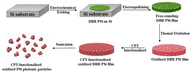
Schematic illustration of the fabrication of CPT-functionalized oxidized porous silicon photonic particles. DBR PSi was prepared by electrochemical etching, detached from the Si substrate by electropolishing, thermally oxidized, functionalized with CPT, and subsequently fragmented by sonication to form photonic particles.
The morphology of the fabricated DBR PSi was examined by FE-SEM, as shown in Fig. 2. The cross-sectional image (Fig. 2(a)) reveals a well-defined multilayer structure extending throughout the PSi film. The periodic contrast observed along the depth direction is attributed to alternating porous layers formed by the periodic modulation of current density during electrochemical etching. This regular layered morphology confirms the successful formation of the DBR structure, in which variations in porosity produce alternating effective refractive indices required for the characteristic photonic response. The surface image (Fig. 2(b)) shows a highly porous and interconnected morphology with nanoscale pores distributed across the PSi surface. Such a porous framework provides a large accessible surface area and internal pore volume for subsequent surface oxidation and CPT functionalization. More importantly, molecules incorporated into or released from these pores can alter the effective refractive index of the porous medium. Therefore, the combination of the periodic multilayer architecture and the accessible porous network provides the structural basis for converting changes associated with CPT release into variations in the optical reflection response. Overall, the SEM observations demonstrate that the electrochemical etching process successfully produced DBR PSi with both a periodically modulated multilayer structure and a highly porous surface morphology. These structural characteristics are essential for the subsequent preparation of oxidized PSi photonic particles and their application in optical monitoring of drug release.
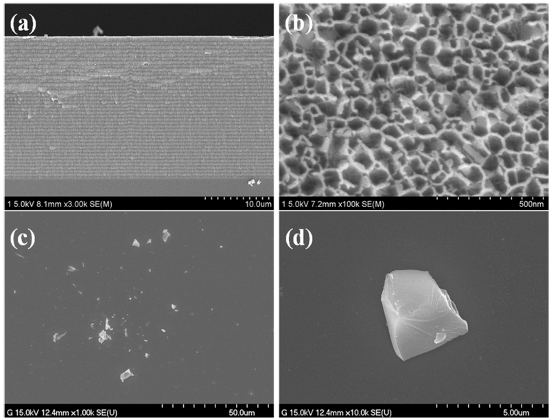
FE-SEM images of DBR porous silicon. (a) cross-sectional morphology showing the periodic multilayer structure, (b) surface morphology showing the porous network, (c) particle distribution after ultrasonic fragmentation, and (d) representative morphology of the fragmented photonic particles.
Following ultrasonic fragmentation, the freestanding CPT-functionalized DBR PSi films were converted into irregularly shaped photonic particles, as shown in Fig. 2(c) and (d). The particle-distribution image (Fig. 2(c)) shows a heterogeneous population of plate-like fragments with lateral dimensions predominantly in the micrometer range, while the representative particle morphology (Fig. 2(d)) shows the presence of smaller fragments within the micrometer-scale population. The broad size distribution is consistent with the mechanical fragmentation of freestanding porous films during ultrasonication. The fragmented particles retain morphological features derived from the porous parent DBR film, supporting their subsequent use in CPT release and optical monitoring experiments. The micrometer-scale and heterogeneous particle size suggests that the present system is more appropriately considered a proof-of-concept platform for localized drug release and optical monitoring rather than a size-controlled carrier for systemic delivery. Further optimization of the fragmentation and size-selection processes would be required for biomedical applications in which a narrowly defined particle size is important.
3.2 Surface Modification and CPT Functionalization
The successive surface modification of DBR PSi was characterized by diffuse-reflectance FT-IR spectroscopy, as shown in Fig. 3. Freshly etched DBR PSi exhibited characteristic Si–H-related absorption bands, including a stretching vibration near 2100 cm-1 and a bending vibration around 900 cm-1, consistent with the hydrogen-terminated surface of electrochemically etched PSi. Following thermal oxidation, the intensity of the Si–H-related bands decreased substantially, while a strong and broad Si–O–Si absorption band developed in the 1000–1250 cm-1 region. Additional oxygen-back-bonded silicon hydride features were observed around 2300 and 850 cm-1. These spectral changes indicate the formation of an oxidized surface while retaining the porous DBR framework.
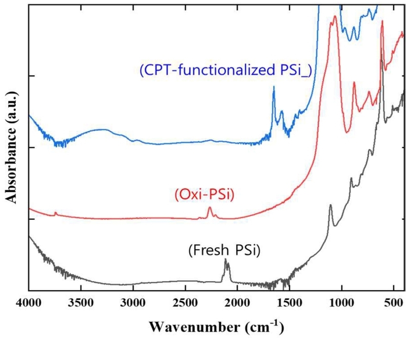
FT-IR spectra of DBR porous silicon at different stages of surface modification: freshly etched DBR PSi, thermally oxidized DBR PSi, and CPT-functionalized oxidized DBR PSi
Following CPT functionalization, additional absorption features were observed in the 1500–1700 cm-1 region compared with those of oxidized DBR PSi. In particular, the features around 1650 and 1570 cm-1 are consistent with vibrational modes associated with CPT, supporting the presence of CPT on the oxidized PSi surface. Together with the pronounced Si–O–Si absorption of the oxidized sample, these changes demonstrate the successive modification of DBR PSi from the freshly etched state to oxidized DBR PSi and subsequently to CPT-functionalized oxidized DBR PSi.
3.3 Optical Response during CPT Release
The optical response of the CPT-functionalized oxidized PSi photonic particles was monitored during CPT release. Fig. 4 shows the time-dependent reflectivity spectra recorded over 12 h. As the release proceeded, the reflection band gradually shifted toward shorter wavelengths, accompanied by a progressive decrease in reflectivity intensity. The blue shift is consistent with a reduction in the effective refractive index of the porous matrix as CPT is released from the pores and surface of the oxidized PSi photonic particles. Despite these spectral changes, the characteristic reflection band remained distinguishable throughout the measurement period, suggesting that the photonic characteristics of the DBR structure were largely retained during CPT release.
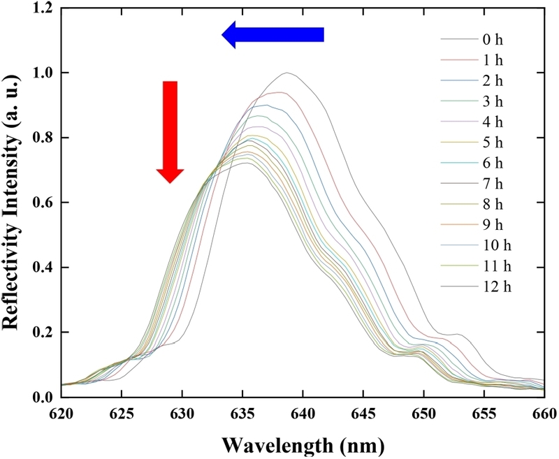
Time-dependent reflectivity spectra of CPT-functionalized oxidized PSi photonic particles during CPT release over 12 h, showing a progressive blue shift and decrease in reflectivity intensity.
3.4 Differential Spectral Analysis and Optical Monitoring of CPT Release
Although the time-dependent reflectivity spectra showed a gradual blue shift during CPT release, the magnitude of the spectral displacement was relatively small. Therefore, direct comparison of the reflectivity spectra, particularly during the early stage of release, did not clearly reveal the small wavelength change. To more clearly distinguish these subtle spectral variations, differential spectral analysis was applied using the initial reflectivity spectrum as a reference. Fig. 5(a) compares the reflectivity spectra measured at 0 and 1 h. The two reflection bands largely overlap, indicating that the spectral change during the initial stage of CPT release is relatively small. However, subtraction of the two spectra produces a distinct differential response, as shown in Fig. 5(c). The characteristic positive and negative features in the differential spectrum arise from the displacement of the reflection band and therefore provide a more readily distinguishable representation of the small wavelength shift than direct comparison of the original spectra. A more pronounced change was observed after 12 h of release. As shown in Fig. 5(b), the reflection band exhibited a clearer blue shift together with a decrease in reflectivity intensity compared with the initial spectrum. Correspondingly, the differential spectrum in Fig. 5(d) showed a larger response than that observed at 1 h. This increase in the differential response is consistent with the progressive change in the optical properties of the porous structure during CPT release. These results indicate that differential spectral analysis provides a useful means of highlighting small release-induced changes in the DBR reflection band. In particular, the differential representation allows subtle spectral displacement to be distinguished even when the original reflection bands substantially overlap. The differential response therefore complements direct analysis of the reflection wavelength and intensity for monitoring the optical changes associated with CPT release.
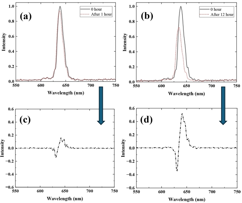
Reflectivity spectra and corresponding differential responses of CPT-functionalized oxidized PSi photonic particles during CPT release. (a) reflectivity spectra at 0 and 1 h, (b) reflectivity spectra at 0 and 12 h, (c) differential spectrum obtained from the 0 and 1 h spectra, and (d) differential spectrum obtained from the 0 and 12 h spectra.
The relationship between CPT release and the optical response of the oxidized PSi photonic particles is shown in Fig. 6. The cumulative percentage of released CPT increased rapidly during the initial and intermediate stages of the experiment and gradually approached a plateau toward 12 h. In contrast, the normalized reflectivity intensity decreased continuously with increasing release time. The opposing trends demonstrate that the optical response of the oxidized PSi photonic particles evolves concurrently with CPT release. The relatively rapid optical change during the early release period followed by a progressively slower response is also consistent with the temporal release profile. The decrease in reflectivity intensity and the blue shift of the reflection band provide complementary information on the release process. The wavelength shift reflects changes in the effective refractive index within the porous photonic structure, whereas the intensity variation provides an additional measure of the evolving optical characteristics of the particles. Together with the differential spectral response, these results demonstrate that CPT release can be monitored through changes in the intrinsic photonic response of the oxidized PSi photonic particles. Overall, the results demonstrate that CPT-functionalized oxidized PSi photonic particles retain their characteristic photonic response during drug release. CPT release induces a systematic blue shift and decrease in reflectivity intensity, while differential spectral analysis enables subtle spectral changes to be more clearly resolved. The corresponding changes in CPT release and optical response indicate that the photonic particles can serve not only as drug carriers but also as optically readable materials for monitoring the release process. These characteristics demonstrate the potential of oxidized PSi photonic particles as a platform integrating drug delivery with intrinsic optical monitoring.
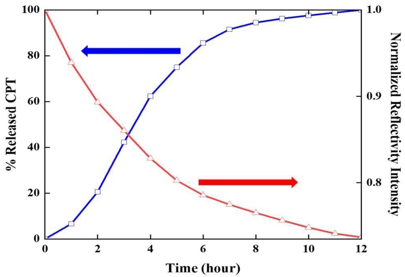
Time-dependent changes in cumulative CPT release and normalized reflectivity intensity of CPT-functionalized oxidized PSi photonic particles.
The intrinsic optical response of DBR PSi photonic particles offers a potential self-reporting strategy for monitoring drug release without the need for additional fluorescent labels or separate sensing components. Porous silicon has been widely investigated as a drug-delivery material because its porous structure, surface chemistry, and optical properties can be tailored for molecular loading, release, and monitoring [6]. In particular, porous silicon photonic crystals have been investigated as potential self-reporting platforms for localized biomedical applications, including intraocular drug delivery [18]. More recently, self-reporting porous silicon microparticles have also been demonstrated, in which intrinsic optical signals were correlated with payload release and degradation of the porous matrix [19]. Based on these previous studies and the present results, the CPT-functionalized DBR PSi particles investigated here are primarily intended as a proof-of-concept platform for localized drug release combined with intrinsic optical monitoring. In future biomedical applications, changes in the characteristic reflectivity of the photonic particles could provide a label-free optical readout of drug-release behavior in locally administered or optically accessible delivery systems. Further optimization of particle size, surface functionalization, and optical readout will be required for practical in vitro or in vivo implementation.
4. CONCLUSIONS
Porous silicon photonic particles based on a DBR structure were successfully prepared for optical monitoring of drug release. The periodic multilayer structure and porous morphology of the fabricated DBR PSi were confirmed by FE-SEM. Following thermal oxidation, the oxidized PSi was functionalized with CPT and subsequently fragmented by sonication to produce photonic particles. The resulting particles exhibited irregular micrometer-scale morphologies while maintaining optical characteristics suitable for monitoring changes associated with CPT release.
CPT release from the photonic particles was evaluated by UV-visible spectroscopy, while the corresponding time-dependent changes in optical reflectivity were simultaneously monitored. As CPT was released from the porous structure, the reflection band shifted toward shorter wavelengths and decreased in intensity, consistent with changes in the effective refractive index of the porous medium. Because the release-associated wavelength shift was relatively small, differential spectral analysis was employed to enhance subtle spectral variations and thereby improve the distinction of the optical response associated with CPT release.
These results demonstrate the feasibility of combining molecular release with an intrinsic optical readout using oxidized PSi photonic particles. The present system is therefore considered primarily as a proof-of-concept platform for localized drug release combined with optical monitoring. In future biomedical applications, the characteristic reflectivity changes of the photonic particles could potentially provide a label-free means of tracking drug-release behavior in locally administered or optically accessible delivery systems, without requiring additional fluorescent labels or separate sensing components. Further optimization of particle size and size distribution, surface functionalization, release characteristics, and optical readout will be necessary to extend this concept toward practical in vitro or in vivo biomedical applications.
REFERENCES
-
A.G. Cullis, L.T. Canham, Visible light emission due to quantum size effects in highly porous crystalline silicon, Nature 353 (1991) 335–338.
[https://doi.org/10.1038/353335a0]

-
L.T. Canham, Bioactive silicon structure fabrication through nanoetching techniques, Adv. Mater. 7 (1995) 1033–1037.
[https://doi.org/10.1002/adma.19950071215]
-
D.B. Mawhinney, J.A. Glass, J.T. Yates Jr., FTIR study of the oxidation of porous silicon, J. Phys. Chem. B 101 (1997) 1202–1206.
[https://doi.org/10.1021/jp963322r]
-
C.A. Prestidge, T.J. Barnes, C.-H. Lau, C. Barnett, A. Loni, L. Canham, Mesoporous silicon: a platform for the delivery of therapeutics, Expert Opin. Drug Deliv. 4 (2007) 101–110.
[https://doi.org/10.1517/17425247.4.2.101]
-
J. Salonen, A.M. Kaukonen, J. Hirvonen, V.-P. Lehto, Mesoporous silicon in drug delivery applications, J. Pharm. Sci. 97 (2008) 632–653.
[https://doi.org/10.1002/jps.20999]
-
E.J. Anglin, L. Cheng, W.R. Freeman, M.J. Sailor, Porous silicon in drug delivery devices and materials, Adv. Drug Deliv. Rev. 60 (2008) 1266–1277.
[https://doi.org/10.1016/j.addr.2008.03.017]
-
V.S.-Y. Lin, K. Motesharei, K.P.S. Dancil, M.J. Sailor, M.R. Ghadiri, A porous silicon-based optical interferometric biosensor, Science 278 (1997) 840–843.
[https://doi.org/10.1126/science.278.5339.840]
-
K.-P.S. Dancil, D.P. Greiner, M.J. Sailor, A porous silicon optical biosensor: detection of reversible binding of IgG to a protein A-modified surface, J. Am. Chem. Soc. 121 (1999) 7925–7930.
[https://doi.org/10.1021/ja991421n]
-
Y.Y. Li, F. Cunin, J.R. Link, T. Gao, R.E. Betts, S.H. Reiver, et al., Polymer replicas of photonic porous silicon for sensing and drug delivery applications, Science 299 (2003) 2045–2047.
[https://doi.org/10.1126/science.1081298]
-
W. Theiß, Optical properties of porous silicon, Surf. Sci. Rep. 29 (1997) 91–192.
[https://doi.org/10.1016/S0167-5729(96)00012-X]
-
T.J. Barnes, K.L. Jarvis, C.A. Prestidge, Recent advances in porous silicon technology for drug delivery, Ther. Deliv. 4 (2013) 811–823.
[https://doi.org/10.4155/tde.13.52]
-
D.J. Savage, X. Liu, S.A. Curley, M. Ferrari, R.E. Serda, Porous silicon advances in drug delivery and immunotherapy, Curr. Opin. Pharmacol. 13 (2013) 834–841.
[https://doi.org/10.1016/j.coph.2013.06.006]
-
T. Kumeria, S.J.P. McInnes, S. Maher, A. Santos, Porous silicon for drug delivery applications and theranostics: recent advances, critical review and perspectives, Expert Opin. Drug Deliv. 14 (2017) 1407–1422.
[https://doi.org/10.1080/17425247.2017.1317245]
-
J.-H. Park, L. Gu, G. von Maltzahn, E. Ruoslahti, S.N. Bhatia, M.J. Sailor, Biodegradable luminescent porous silicon nanoparticles for in vivo applications, Nat. Mater. 8 (2009) 331–336.
[https://doi.org/10.1038/nmat2398]
-
T. Tanaka, L.S. Mangala, P.E. Vivas-Mejia, R. Nieves-Alicea, A.P. Mann, E. Mora, et al., Sustained small interfering RNA delivery by mesoporous silicon particles, Cancer Res. 70 (2010) 3687–3696.
[https://doi.org/10.1158/0008-5472.CAN-09-3931]
-
A. Tzur-Balter, Z. Shatsberg, M. Beckerman, E. Segal, N. Artzi, Mechanism of erosion of nanostructured porous silicon drug carriers in neoplastic tissues, Nat. Commun. 6 (2015) 6208.
[https://doi.org/10.1038/ncomms7208]
-
E.J. Anglin, M.P. Schwartz, V.P. Ng, L.A. Perelman, M.J. Sailor, Engineering the chemistry and nanostructure of porous silicon Fabry–Pérot films for loading and release of a steroid, Langmuir 20 (2004) 11264–11269.
[https://doi.org/10.1021/la048105t]
-
L. Cheng, E. Anglin, F. Cunin, D. Kim, M.J. Sailor, I. Falkenstein, et al., Intravitreal properties of porous silicon photonic crystals: a potential self-reporting intraocular drug-delivery vehicle, Br. J. Ophthalmol. 92 (2008) 705–711.
[https://doi.org/10.1136/bjo.2007.133587]
-
J. Wang, T. Kumeria, M.T. Bezem, J. Wang, M.J. Sailor, Self-reporting photoluminescent porous silicon microparticles for drug delivery, ACS Appl. Mater. Interfaces 10 (2018) 3200–3209.
[https://doi.org/10.1021/acsami.7b09071]
-
S.J.P. McInnes, E.J. Szili, S.A. Al-Bataineh, J. Xu, M.E. Alf, K.K. Gleason, et al., Combination of iCVD and porous silicon for the development of a controlled drug delivery system, ACS Appl. Mater. Interfaces 4 (2012) 3566–3574.
[https://doi.org/10.1021/am300621k]