
Dynamic-Mode Microcantilever Biosensor: Detection Strategies and Recent Applications
ⓒ The Korean Sensors Society
This is an Open Access article distributed under the terms of the Creative Commons Attribution Non-Commercial License (https://creativecommons.org/licenses/by-nc/3.0/) which permits unrestricted non-commercial use, distribution, and reproduction in any medium, provided the original work is properly cited.
Abstract
Microcantilever biosensors have attracted considerable attention as micro-electro-mechanical system (MEMS)-based sensing platforms because of their high mechanical sensitivity, label-free operation, and real-time detection capability. By directly converting biomolecular interactions into mechanical responses through surface stress or resonance-frequency shifts, microcantilever sensors enable highly sensitive, quantitative analysis without requiring fluorescent or enzymatic labels. This review summarizes recent advances in dynamic-mode microcantilever biosensors, focusing on their operating principles, excitation methods, and signal transduction techniques. Representative applications in nucleic acid, protein, and cell detection are discussed, highlighting recent developments in molecular recognition strategies and nanomaterial-assisted signal amplification. These advances have significantly improved analytical performance, enabling ultrasensitive detection from single-copy nucleic acids to picogram-level proteins and single-cell analysis. Despite this progress, several technical challenges still limit the practical application of microcantilever biosensors. Future advances in portable, integrated readout technologies; standardized, scalable fabrication; and multiplexed sensing are expected to facilitate the translation of microcantilever biosensors from laboratory-scale demonstrations to practical applications in clinical diagnostics, environmental monitoring, and food safety.
Keywords:
Microcantilever, Biosensor, Dynamic mode, Nucleic acid detection, Protein detection, Cell detection1. INTRODUCTION
Cantilever structures have a long history in engineering, and cantilever beams have been widely used in mechanical and civil engineering because of their simple geometry and excellent load-bearing characteristics [1]. The rapid advancement of micro and nanofabrication technologies has profoundly influenced the development of mechanical structures with dimensions down to a few nanometers, allowing unprecedented control over geometry, mass, and mechanical properties [2]. Microcantilever structures have become attractive sensing platforms because of their simple fabrication process, high mechanical sensitivity, low power consumption, and compatibility with label-free detection [3]. Furthermore, microcantilever sensors can detect extremely small forces, mass loading, and surface stress variations through static deflection or resonance frequency shifts [4]. Consequently, microcantilever sensors have established themselves as one of the most sensitive and versatile sensing platforms among existing sensor technologies [4].
A major advantage of microcantilever sensors is their ability to directly detect interaction forces between target analytes and the functionalized microcantilever surface [5]. Unlike electrical or optical sensors that primarily detect secondary effects such as charge transfer or refractive index changes, microcantilever sensors can directly measure surface stress and binding forces arising from molecular interactions. Recent force-sensitive cantilever studies have enabled quantitative analysis of intermolecular forces and adsorption-induced stress [6-12].
Microcantilever sensors can be operated in two modes: static and dynamic. In static mode, detection is based on surface stress-induced deflection, which is often disturbed by environmental fluctuations such as nonspecific adsorption and long-term thermal drift [4,5,13]. In contrast, dynamic-mode sensing transduces biomolecular interactions into resonance frequency shifts, enabling direct, quantitative detection of mass loading with significantly improved signal-to-noise ratios. The signal from a dynamic-mode microcantilever sensor is less affected by low-frequency noise and baseline drift, allowing stable and reproducible measurements compared with static mode [4,5]. Consequently, dynamic-mode operation has become a preferred sensing strategy for microcantilever-based biosensors because of its sensitivity, robustness, and quantitative reliability [5].
Microcantilever sensors can detect a wide range of biological species, from small biomolecules to whole cells. By functionalizing the cantilever surface with specific recognition elements such as DNA probes, antibodies, or aptamers, the sensors can selectively detect nucleic acids (DNA and RNA) [14-16], proteins [17-21], and even single cells [22-24]. The microcantilever sensors can measure simple presence or concentration, like other types of sensors. However, the sensors are also able to measure surface stress, binding-induced mass loading, and viscoelastic or mechanical property changes associated with biomolecular interactions, which could not be achieved with other types of sensors such as electrical and optical sensors [25]. This unique detection capability enables microcantilever-based platforms to perform biosensing across molecular-to-cellular scales, highlighting their potential for diverse biosensing applications.
In this review, we provide a comprehensive overview of recent researches of microcantilever-based biosensors published after 2020, focusing on detection strategies and representative biological applications. First, we describe the fundamental operating principles of dynamic sensing modes, followed by a discussion of mechanical excitation methods and signal transduction techniques. Finally, we discuss recent applications of microcantilever biosensors and current challenges, offering perspectives on future directions in biomedical diagnostics, environmental monitoring, and fundamental biological studies.
2. DETECTION STRATEGIES
2.1 Detection Mechanism
The resonance frequency of a microcantilever is a fundamental mechanical property determined primarily by its effective stiffness and effective mass [26]. For a simple rectangular cantilever beam with small thickness, the resonance frequency can be approximated using the classical elastic continuum model as below:
| (1) |
where fₙ is the resonance frequency of the nth mode, λₙ is the dimensionless eigenvalue determined by the boundary conditions, L, E, I, ρ, and A are the length, Young's modulus, cross-sectional moment of inertia, mass density, and cross-sectional area of the microcantilever, respectively. Eq. (1) can be simplified using the effective stiffness and effective mass as below:
| (2) |
where f₀ is the fundamental resonance frequency, keff is the effective stiffness, and meff is the effective mass of the cantilever. From the equation, the resonance frequency increases with greater stiffness and decreases with increasing mass. In biosensing applications, this dependence underpins the detection mechanism of a dynamic-mode sensor. For instance, when target analytes bind to the surface of the cantilever, the additional mass leads to a measurable decreased resonance frequency (Fig. 1). Specifically, the resonance frequency scales inversely with the cantilever length and directly with its thickness, indicating that shorter cantilevers can achieve higher resonance frequencies [27]. Reducing the cantilever dimensions can also improve mass sensitivity by decreasing its effective mass. Material properties such as density and elastic modulus also influence the cantilever's dynamic behavior. However, these properties are generally fixed because microcantilevers are predominantly fabricated from silicon and silicon nitride using semiconductor manufacturing processes, as well as polymers, piezoelectric materials, and diamond-based materials compatible with semiconductor manufacturing processes.
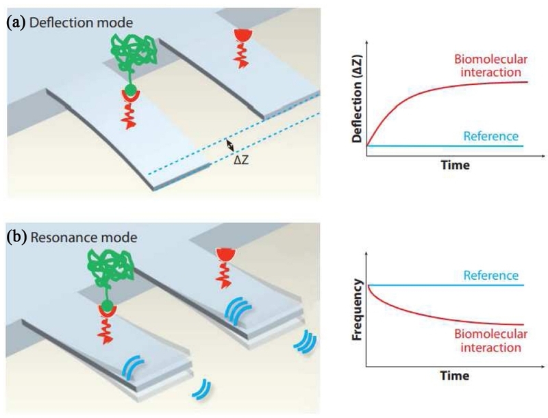
Two modes of microcantilever based biomolecule detection. (a) deflection(static) mode. (b) dynamic mode. Adapted from Ref. [13].
In dynamic-mode biosensing, resonance frequency serves as the primary sensing parameter for detection in response to mass loading on the cantilever surface. When target analytes bind to receptors immobilized on the cantilever, the effective mass increases, resulting in a measurable frequency shift that can be correlated quantitatively with the amount of bound analyte. Beyond simple mass effects, the viscoelastic properties of the adsorbed biological layer can also modify effective stiffness and damping, influencing resonance behavior [4,27].
2.2 Mechanical Excitation Method
The resonance frequency of a microcantilever is measured with controlled excitation of the cantilever and monitoring of its vibrational response. Several actuation methods are commonly employed. Piezoelectric base excitation is one of the simplest techniques, in which a piezoelectric actuator mechanically vibrates the cantilever substrate, as shown in Fig. 2 [28,30]. The substrate then transfers energy to the cantilever, causing it to oscillate near its resonance frequency. A major advantage of this method is its ease of implementation. However, this method may introduce additional substrate resonances in liquid environments, affecting the detection signal.
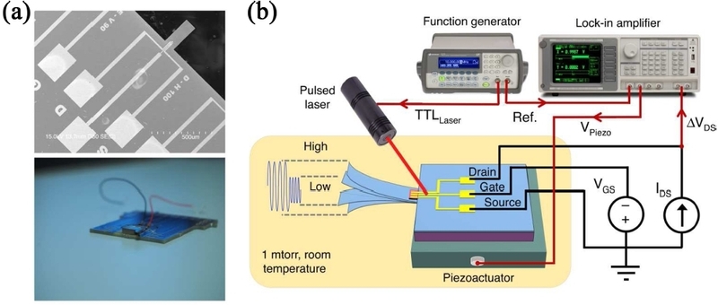
Microcantilever excitation using piezoelectric base. (a) SEM image of the microcantilever (top) and photo of the piezo actuator attached underneath the microcantilever chip (bottom). (b) Schematic diagram of the experimental setup showing simultaneous piezo chip and photoacoustic excitation. Adapted from Ref. [28].
Electrothermal actuation relies on periodic Joule heating of an integrated resistive element within or near the cantilever [30]. The resulting thermal expansion generates bending oscillations (Fig. 3). This approach allows compact on-chip integration but may induce thermal drift, requiring careful experimental preparation and control. Magnetic actuation is achieved by coating the cantilever with a thin magnetic layer and applying an external oscillating magnetic field [31]. In detail, the magnetic torque drives cantilever oscillation without mechanically disturbing the substrate, improving performance in liquid media. The electrostatic technique uses an applied AC voltage between the cantilever and a nearby electrode to generate an oscillatory electrostatic force [32,33]. This method is widely used in micro-electro-mechanical systems(MEMS) due to its low power consumption and compatibility with electronic circuit integration. Photothermal excitation employs a modulated laser beam to periodically heat the cantilever surface, inducing localized thermal expansion and vibration. This technique provides resonance excitation with minimal mechanical coupling to the environment and is particularly advantageous for liquid-phase biosensing.
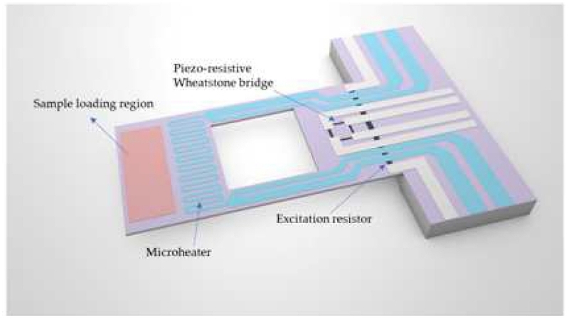
Schematic diagram of a microcantilever excitation using an electrothermal actuation method. Adapted from Ref. [30].
2.3 Mechanical Response Measurement
For microcantilever sensors, the mechanical response readout mechanism converts the mechanical response into measurable electrical or optical signals, enabling precise measurement of resonance frequency in each state. Readout performance influences sensor characteristics such as sensitivity, resolution, and system integration. Among the various techniques, optical and electrical transduction are the most commonly used approaches. Optical beam deflection is a standard approach in laboratory-based systems [34]. In this configuration, a laser beam is focused onto the edge of the cantilever, and a position-sensitive photodetector detects the reflected beam (Fig. 4). Mechanical motion induces angular deflection of the reflected beam, which the photodetector converts into a voltage signal proportional to cantilever displacement. This technique provides high displacement sensitivity and wide bandwidth, although it requires precise optical alignment and external instrumentation.
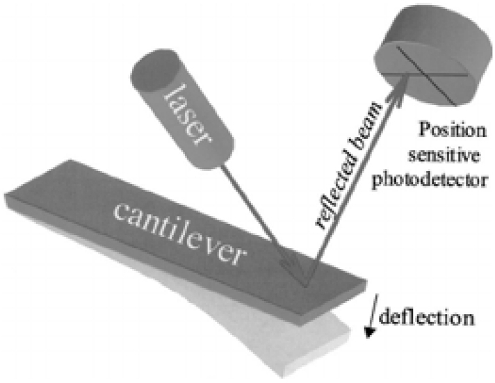
Schematic diagram of a microcantilever mechanical response measurement using an optical method. Adapted from Ref. [34].
Laser Doppler vibrometry (LDV) is another optical readout technique that measures cantilever vibration based on the Doppler frequency shift of the reflected laser beam. This non-contact method provides highly sensitive vibration measurements and enables accurate determination of the resonance frequency. Electrical transduction methods convert microcantilever response into electrical signals. Common approaches include piezoresistive, piezoelectric, and capacitive-based methods [35-37]. Piezoresistive methods detect strain-induced resistance changes, piezoelectric methods generate electrical charges during deformation, and capacitive methods measure changes in electrode capacitance (Fig. 5). These methods offer advantages such as miniaturization, low-cost semiconductor integration, array compatibility, and portability compared with conventional optical readout systems. However, thermal drift, electrical noise, parasitic capacitance, and electromagnetic interference can degrade detection performance.
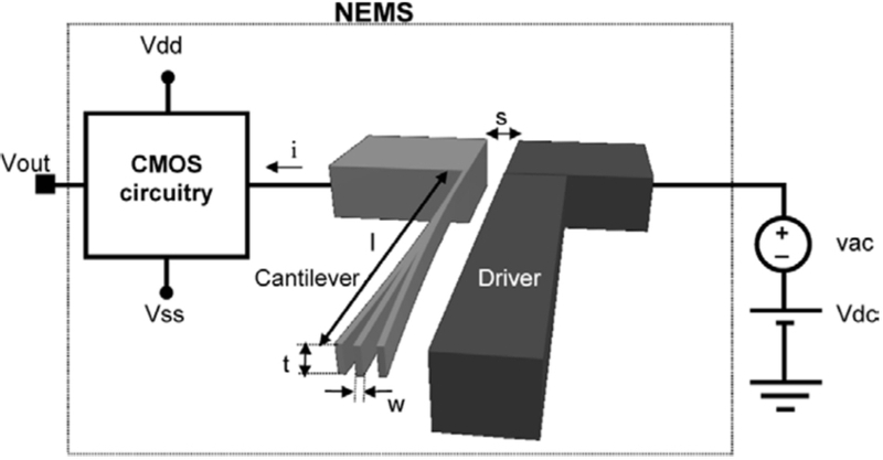
Schematic diagram of a microcantilever mechanical response measurement using a capacitive method. Adapted from Ref. [37].
3. RECENT RESEARCH
Microcantilever biosensors have emerged as one of the most promising MEMS-enabled sensing platforms for the label-free, highly sensitive detection of biological species. Fritz et al. first demonstrated a microcantilever biosensor in 2000, translating DNA hybridization into measurable nanomechanical surface stress and establishing the foundation for label-free microcantilever biosensing [38]. Since then, microcantilever biosensors have evolved considerably, enabling sensitive, label-free detection of diverse biological targets, including nucleic acids, proteins, and cells. More recently, researchers have combined microcantilever biosensors with nanomaterials such as graphene, carbon nanotubes, and gold nanoparticles to increase surface area and amplify signals [39]. Integration with molecular recognition systems has further enhanced sequence specificity and enabled ultrasensitive detection of biological targets.
Microcantilever biosensors have demonstrated excellent potential as nucleic acid detection platforms. Lei et al. developed a dynamic-mode microcantilever integrated with recombinase polymerase amplification (RPA) and gold nanoparticles (AuNPs) for ultrasensitive detection of the plant pathogen Leptosphaeria maculans, as shown in Fig. 6 [40]. Unlike conventional biosensors integrated with nucleic acid assays such as polymerase chain reaction (PCR) that rely on optical or electrochemical labels, microcantilever sensors directly convert biomolecular recognition events into measurable mechanical signals. Although the platform supports both static and dynamic operation, the dynamic mode was employed as the primary sensing mechanism by monitoring resonance frequency shifts induced by mass loading and stiffness changes after target binding. The cantilever was actively excited using a piezoelectric crystal, while the optical readout system tracked the cantilever motion. Sequential binding of biotin-labeled RPA amplicons and complementary DNA-functionalized 20 nm AuNPs significantly amplified the resonance frequency shift, leading to a detection limit of approximately one copy of genomic DNA, which is more sensitive than conventional fluorescence RPA. The assay also detected 57 ppm of L. maculans genomic DNA in field samples, demonstrating excellent sensitivity, specificity, and suitability for rapid on-site plant pathogen diagnostics.
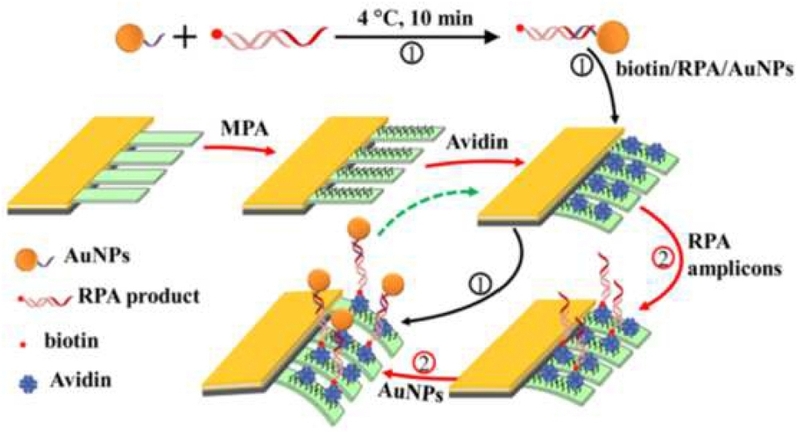
Schematic diagram of DNA detection using microcantilever with AuNPs. Adapted from Ref. [40].
Microcantilever biosensors have demonstrated excellent performance for protein biomarker detection. Zhao et al. developed a dynamic-mode microcantilever using AuNPs as a mass amplifier for highly sensitive detection of the protein biomarker alpha-fetoprotein (AFP) [41]. The sensor operated with a piezoelectric transducer for mechanical excitation under vacuum. The optical system measured the mechanical response. For detection, antibody probes were functionalized on the microcantilever surface to bind AFP specifically (Fig. 7). AuNP-conjugated detection antibodies were then introduced to form a sandwich immunoassay. The AuNP-assisted sandwich immunoassay enhanced the resonance frequency response from approximately 2 Hz to 140 Hz for 20 ng/mL AFP, increasing the sensor sensitivity from 7.68 Hz/nM to 412 Hz/nM and reducing the limit of detection (LOD) from 1.1 ng/mL to 21 pg/mL. Integrating microcantilevers with AuNP-based sandwich immunoassays dramatically enhanced mass-loading sensitivity while maintaining high reproducibility, demonstrating strong potential for early clinical diagnosis of protein biomarkers.
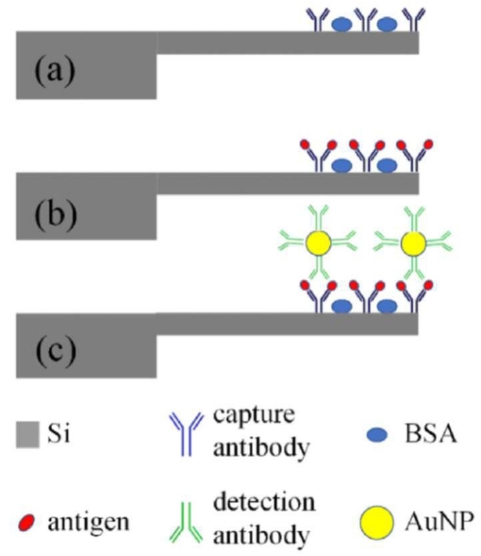
Schematic diagram of protein biomarker detection using a microcantilever. (a) Microcantilever surface functionalization using capture antibodies and BSA. (b) Specific binding of target antigens to the capture antibodies. (c) AuNP-assisted target detection for signal amplification. Adapted from Ref. [41].
Microcantilever biosensors have demonstrated outstanding capabilities for cell detection by combining label-free sensing, high sensitivity, real-time monitoring, and compatibility with microfluidic and array-based platforms. Incaviglia et al. optimized microcantilevers for single adherent mammalian cell mass measurements by reducing cantilever inertial mass through focused ion beam (FIB) modification, as shown in Fig. 8 [42]. The redesigned cantilevers exhibited over 70% improvement in signal-to-noise ratio and picogram mass sensitivity, with significantly reduced measurement errors caused by cell migration. Fathy and Lai developed a dynamic V-shaped microcantilever integrated with dielectrophoretic (DEP) particle enrichment for real-time E. coli detection [43]. The sensor detected bacterial concentrations ranging from 10³ to 10⁵ cells/mL within 4 min, producing frequency shifts of −755 to −2264 Hz. More recently, Qiao et al. introduced a graphene oxide-assisted magnetic nanomechanical microcantilever array that substantially improved mechanical signal amplification [44]. The platform achieved single bacterial cell detection without culture, maintained a linear detection range of 1–100 CFU/mL, enabled simultaneous detection of multiple bacterial species, and demonstrated excellent specificity, reproducibility, stability, and resistance to interference in complex samples such as milk.
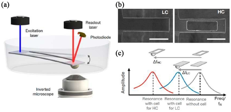
Microcantilever for cell detection. (a) Schematic diagram of cell detection using microcantilever. (b) Top view SEM images of rectangular (LC) and FIB-modified (HC) microcantilevers. Scale bars, 50 μm. (c) The resonance frequency shift of each microcantilever state upon cell attachment. Adapted from Ref. [42].
4. CHALLENGES AND PERSPECTIVES
Despite their high sensitivity and label-free detection capability, microcantilever biosensors have not achieved widespread adoption comparable to mature MEMS devices such as accelerometers. One major limitation, particularly for dynamic-mode sensing, is operation in liquid environments. Biological reactions generally require aqueous conditions. The surrounding liquid introduces hydrodynamic added mass and viscous damping, which decrease the resonance frequency, broaden the resonance peak, and consequently reduce frequency resolution and mass sensitivity [45]. Furthermore, variations in temperature, fluid density, and viscosity can cause resonance-frequency drift independent of target binding, resulting in false or nonspecific sensor responses. However, differential measurements using reference cantilevers and temperature compensation can reduce these effects, although these approaches increase system complexity.
The intrinsic mechanical response generated by biomolecular binding can be extremely small, particularly for targets with low molecular weight and low target concentrations. Recent studies have therefore increasingly employed mass amplification strategies using AuNPs, enabling approximately 70-fold improvement in the detection limit for AFP (Table 1) and significantly enhanced measurement reproducibility by increasing the specific signal above thermal noise and nonspecific adsorption [41]. Similarly, combining RPA with AuNP-assisted mass loading has enabled nucleic acid detection down to approximately a single DNA copy [40]. These approaches demonstrate the exceptional sensitivity achievable by microcantilever platforms; however, they also introduce additional labeling, reaction, and sample-preparation steps, partially compromising the inherent simplicity and label-free nature of mechanical sensing. Therefore, alternative amplification strategies that maintain simple assay procedures while ensuring sufficiently strong and reproducible mechanical responses are required.

Comparison of sensor performance of representative dynamic-mode microcantilever biosensors.
Furthermore, conventional optical readout requires bulky instrumentation and precise alignment, limiting system miniaturization [34]. Future development should therefore focus on standardized, scalable MEMS fabrication; robust surface functionalization; integrated readout and microfluidics; and multiplexed cantilever arrays. Greater validation using complex real-world samples will also be essential to translate microcantilever biosensors into reliable clinical diagnostics, environmental monitoring, and food safety applications.
5. CONCLUSIONS
Microcantilever biosensors have emerged as highly sensitive and versatile MEMS-based sensing platforms that translate biomolecular interactions into measurable mechanical signals. Recent advances in dynamic-mode sensing and signal transduction have significantly improved the detection of diverse biological targets, including nucleic acids, proteins, and cells, enabling rapid, real-time, and quantitative analysis with high sensitivity and specificity. In particular, incorporating nanomaterials, advanced molecular recognition strategies, and array-based configurations has substantially enhanced analytical performance. Despite these advances, several challenges remain for practical implementation, including performance degradation in liquid environments, weak mechanical responses to low-mass targets, and limitations in system integration and standardization. Signal amplification, surface functionalization, and microfluidic integration have addressed some of these limitations; however, further improvements are required to achieve robust, reproducible, and simplified sensing platforms.
In addition, conventional optical readout systems require relatively bulky and expensive instrumentation, limiting portability and point-of-care applications, whereas electrical transduction provides a compact alternative but may suffer from lower sensitivity and electrical noise. Large-scale integration, multiplexed detection, device standardization, and scalable manufacturing also remain important barriers to commercialization. Therefore, future research should focus on improving signal stability and device robustness, developing compact, integrated readout systems, integrating microfluidics and on-chip electronics, and establishing standardized, scalable fabrication processes. Advances in these areas, together with enhanced multiplexing capabilities and validation using complex real-world samples, will be essential for translating microcantilever biosensors from laboratory-scale demonstrations into practical platforms for clinical diagnostics, environmental monitoring, and food safety.
Acknowledgments
This research received no external funding.
REFERENCES
- S.P. Timoshenko, History of Strength of Materials, McGraw-Hill, New York, 1953.
-
A. Boisen, S. Dohn, S.S. Keller, S. Schmid, M. Tenje, Cantilever-like micromechanical sensors, Rep. Prog. Phys. 74 (2011) 036101.
[https://doi.org/10.1088/0034-4885/74/3/036101]

-
J. Fritz, Cantilever biosensors, Analyst 133 (2008) 855–863.
[https://doi.org/10.1039/b718174d]
-
J.L. Arlett, E.B. Myers, M.L. Roukes, Comparative advantages of mechanical biosensors, Nat. Nanotechnol. 6 (2011) 203–215.
[https://doi.org/10.1038/nnano.2011.44]
-
B.N. Johnson, R. Mutharasan, Biosensing using dynamic-mode cantilever sensors: A review, Biosens. Bioelectron. 32 (2012) 1–18.
[https://doi.org/10.1016/j.bios.2011.10.054]
-
D.W. Dareing, T. Thundat, Simulation of adsorption-induced stress of a microcantilever sensor, J. Appl. Phys. 97 (2005) 043526.
[https://doi.org/10.1063/1.1853496]
-
M. Godin, V. Tabard-Cossa, Y. Miyahara, T. Monga, P.J. Williams, L.Y. Beaulieu, et al., Cantilever-based sensing: the origin of surface stress and optimization strategies, Nanotechnology 21 (2010) 075501.
[https://doi.org/10.1088/0957-4484/21/7/075501]
-
Y. Zhang, Determining the adsorption-induced surface stress and mass by measuring the shifts of resonant frequencies, Sens. Actuators A Phys. 194 (2013) 169–175.
[https://doi.org/10.1016/j.sna.2013.01.029]
-
Z. Hu, T. Thundat, R.J. Warmack, Investigation of adsorption and absorption-induced stresses using microcantilever sensors, J. Appl. Phys. 90 (2001) 427–431.
[https://doi.org/10.1063/1.1378333]
-
K.M. Goeders, J.S. Colton, L.A. Bottomley, Microcantilevers: Sensing chemical interactions via mechanical motion, Chem. Rev. 108 (2008) 522–542.
[https://doi.org/10.1021/cr0681041]
-
X. Yi, H.L. Duan, Surface stress induced by interactions of adsorbates and its effect on deformation and frequency of microcantilever sensors, J. Mech. Phys. Solids 57 (2009) 1254–1266.
[https://doi.org/10.1016/j.jmps.2009.04.010]
-
T. Yang, X. Li, Y. Chen, D.-W. Lee, G . Zuo, Adsorption induced surface-stress sensing signal originating from both vertical interface effects and intermolecular lateral interactions, Analyst 136 (2011) 5261–5269.
[https://doi.org/10.1039/c1an15695k]
-
K.S. Hwang, S.M. Lee, S.K. Kim, J.H. Lee, T.S. Kim, Micro- and nanocantilever devices and systems for biomolecule detection, Annu. Rev. Anal. Chem. 2 (2009) 77–98.
[https://doi.org/10.1146/annurev-anchem-060908-155232]
-
J. Mertens, C. Rogero, M. Calleja, D. Ramos, J.A. Martín-Gago, C. Briones, et al., Label-free detection of DNA hybridization based on hydration-induced tension in nucleic acid films, Nat. Nanotechnol. 3 (2008) 301–307.
[https://doi.org/10.1038/nnano.2008.91]
-
S.M. Yang, C. Chang, T.I. Yin, P.L. Kuo, DNA hybridization measurement by self-sensing piezoresistive microcantilevers in CMOS biosensor, Sens. Actuators B Chem. 130 (2008) 674–681.
[https://doi.org/10.1016/j.snb.2007.10.072]
-
H.H. Jung, J.S. Park, J.M. Yang, S.W. Lee, K.H. Eom, T.Y. Kwon, et al., Study on the chemical environment for conformational change of i-motif DNA by atomic force microscopy cantilever, J. Sens. Sci. Technol. 19 (2010) 214–220.
[https://doi.org/10.5369/JSST.2010.19.3.214]
-
G. Wu, R.H. Datar, K.M. Hansen, T. Thundat, R.J. Cote, A. Majumdar, Bioassay of prostate-specific antigen (PSA) using microcantilevers, Nat. Biotechnol. 19 (2001) 856–860.
[https://doi.org/10.1038/nbt0901-856]
-
C.A. Savran, S.M. Knudsen, A.D. Ellington, S.R. Manalis, Micromechanical detection of proteins using aptamer-based receptor molecules, Anal. Chem. 76 (2004) 3194–3198.
[https://doi.org/10.1021/ac049859f]
-
W. Shu, S. Laurenson, T.P.J. Knowles, P.K. Ferrigno, A.A. Seshia, Highly specific label-free protein detection from lysed cells using internally referenced microcantilever sensors, Biosens. Bioelectron. 24 (2008) 233–237.
[https://doi.org/10.1016/j.bios.2008.03.036]
-
M. Yue, J.C. Stachowiak, H. Lin, R. Datar, R.J. Cote, A. Majumdar, Label-Free Protein Recognition Two-Dimensional Array Using Nanomechanical Sensors, Nano Lett. 8 (2008) 520–524.
[https://doi.org/10.1021/nl072740c]
-
J.E. Park, D.S. Kim, H.J. Choi, J.K. Shin, P.K. Kim, G. Lim, Absorption analysis of streptavidin–biotin complexes using AFM, J. Sens. Sci. Technol. 15 (2006) 237–244.
[https://doi.org/10.5369/JSST.2006.15.4.237]
-
X. Chen, Y. Pan, H. Liu, X. Bai, N. Wang, B. Zhang, Label-free detection of liver cancer cells by aptamer-based microcantilever biosensor, Biosens. Bioelectron. 79 (2016) 353–358.
[https://doi.org/10.1016/j.bios.2015.12.060]
-
K. Park, J. Jang, D. Irimia, J. Sturgis, J. Lee, J.P. Robinson, et al., ‘Living cantilever arrays' for characterization of mass of single live cells in fluids, Lab Chip 8 (2008) 1034–1041.
[https://doi.org/10.1039/b803601b]
-
B. Łabędź, A. Wańczyk, Z. Rajfur, Precise mass determination of single cell with cantilever-based microbiosensor system, PLoS One 12 (2017) e0188388.
[https://doi.org/10.1371/journal.pone.0188388]
-
S.J. Hyun, H.S. Kim, Y.J. Kim, H.I. Jung, Rapid detection of liposome by piezoresistive cantilever sensor, J. Sens. Sci. Technol. 14 (2005) 156–159.
[https://doi.org/10.5369/JSST.2005.14.3.156]
-
K. Eom, H.S. Park, D.S. Yoon, T. Kwon, Nanomechanical resonators and their applications in biological/chemical detection: Nanomechanics principles, Phys. Rep. 503 (2011) 115–163.
[https://doi.org/10.1016/j.physrep.2011.03.002]
-
J.D. Parkin, G. Hähner, Mass determination and sensitivity based on resonance frequency changes of the higher flexural modes of cantilever sensors, Rev. Sci. Instrum. 82 (2011) 035108.
[https://doi.org/10.1063/1.3563724]
-
F. Bayram, D. Gajula, D. Khan, G. Koley, Mechanical memory operations in piezotransistive GaN microcantilevers using Au nanoparticle-enhanced photoacoustic excitation, Microsyst. Nanoeng. 8 (2022) 8.
[https://doi.org/10.1038/s41378-021-00330-6]
-
C.I. Kim, J.S. Yun, W.-I. Park, Y.-H. Jeong, Y.W. Hong, J.-H. Cho, et al., Power Generation Characteristics of Unimorph Piezoelectric Cantilever with Different Vibration Angle, J. Sens. Sci. Technol. 26 (2017) 107–113.
[https://doi.org/10.5369/JSST.2017.26.2.107]
-
S. Wang, Z. Cao, X. Zhang, H. Yu, L. Yao, An Engineering Method for Resonant Microcantilever Using Double-Channel Excitation and Signal Acquisition Based on LabVIEW, Micromachines 14 (2023) 823.
[https://doi.org/10.3390/mi14040823]
-
N.N. Gosvami, P.C. Nalam, A.L. Exarhos, Q. Tam, J.M. Kikkawa, R.W. Carpick, Direct torsional actuation of microcantilevers using magnetic excitation, Appl. Phys. Lett. 105 (2014) 093101.
[https://doi.org/10.1063/1.4894737]
-
Y.C. Hu, C.M. Chang, S.C. Huang, Some design considerations on the electrostatically actuated microstructures, Sens. Actuators A Phys. 112 (2004) 155–161.
[https://doi.org/10.1016/j.sna.2003.12.012]
-
J.C. Lee, B.K. Choi, Fabrication and Characterization of Electrostatically Actuated Microcantilever Mass Sensors, J. Sens. Sci. Technol. 20 (2011) 40–45.
[https://doi.org/10.5369/JSST.2011.20.1.40]
-
N.V. Lavrik, M.J. Sepaniak, P.G. Datskos, Cantilever transducers as a platform for chemical and biological sensors, Rev. Sci. Instrum. 75 (2004) 2229–2253.
[https://doi.org/10.1063/1.1763252]
-
Z. Wang, R. Yue, R. Zhang, L. Liu, Design and optimization of laminated piezoresistive microcantilever sensors, Sens. Actuators A Phys. 120 (2005) 325–336.
[https://doi.org/10.1016/j.sna.2004.12.006]
-
M.S.K. Mutyala, D. Bandhanadham, L. Pan, V.R. Pendyala, H.-F. Ji, Mechanical and electronic approaches to improve the sensitivity of microcantilever sensors, Acta Mech. Sin. 25 (2009) 1–12.
[https://doi.org/10.1007/s10409-008-0222-6]
-
J. Verd, G. Abadal, J. Teva, M.V. Gaudó, A. Uranga, X. Borrisé, et al., Design, fabrication, and characterization of a submicroelectromechanical resonator with monolithically integrated CMOS readout circuit, J. Microelectromech. Syst. 14 (2005) 508–519.
[https://doi.org/10.1109/JMEMS.2005.844845]
-
J. Fritz, M.K. Baller, H.P. Lang, H. Rothuizen, P. Vettiger, E. Meyer, et al., Translating biomolecular recognition into nanomechanics, Science 288 (2000) 316–318.
[https://doi.org/10.1126/science.288.5464.316]
-
J. Tamayo, P.M. Kosaka, J.J. Ruz, Á. San Paulo, M. Calleja, Biosensors based on nanomechanical systems, Chem. Soc. Rev. 42 (2013) 1287–1311.
[https://doi.org/10.1039/C2CS35293A]
-
R. Lei, P. Wu, L. Li, Q. Huang, J. Wang, D. Zhang, et al., Ultrasensitive isothermal detection of a plant pathogen by using a gold nanoparticle-enhanced microcantilever sensor, Sens. Actuators B Chem. 338 (2021) 129874.
[https://doi.org/10.1016/j.snb.2021.129874]
-
J. Zhao, L. Wang, D. Fu, D. Zhao, Y. Wang, Q. Yuan, et al., Gold nanoparticles amplified microcantilever biosensor for detecting protein biomarkers with high sensitivity, Sens. Actuators A Phys. 321 (2021) 112563.
[https://doi.org/10.1016/j.sna.2021.112563]
-
I. Incaviglia, S. Herzog, G. Fläschner, N. Strohmeyer, E. Tosoratti, D.J. Müller, Tailoring the sensitivity of microcantilevers to monitor the mass of single adherent living cells, Nano Lett. 23 (2023) 588–596.
[https://doi.org/10.1021/acs.nanolett.2c04198]
-
J. Fathy, Y. Lai, A V-Shaped Microcantilever Sensor Based on a Gap Method for Real-Time Detection of E. coli Bacteria, Biosensors 12 (2022) 194.
[https://doi.org/10.3390/bios12040194]
-
Z. Qiao, K. Mei, Y. Wang, T. Yan, S. Zhang, Q. Chen, Y. Chen, C. Wang, T. Ren, S. Wu, Q. Zhang, Ultra-sensitive magnetic nanomechanical array sensor based on graphene oxide for single bacterial cell detection, Nano Res. 18 (2025) 94907290.
[https://doi.org/10.26599/NR.2025.94907290]
-
J.E. Sader, Frequency response of cantilever beams immersed in viscous fluids with applications to the atomic force microscope, J. Appl. Phys. 84 (1998) 64–76.
[https://doi.org/10.1063/1.368002]

Kuewhan Jang is an Associate Professor in the Department of Mechanical Engineering at Hoseo University (Asan, Republic of Korea). He received his B.S. degree in 2010 and Ph.D. degree in 2015 in mechanical engineering from Korea University (Seoul, Republic of Korea). From 2015 to 2016, he was a research professor at Korea University, and he joined Hoseo University in 2017. His research focuses on micro/nanomechanical biosensors, MEMS/NEMS, microcantilever sensors, nanomaterials, and biosensing technologies. His work integrates MEMS platforms, surface functionalization, and nanomaterials to develop highly sensitive, label-free biosensing platforms for detecting nucleic acids, metal ions, and environmental contaminants. He teaches courses including solid mechanics, engineering materials, sensor engineering, engineering mathematics, mechanical vibrations, and statics.